
Fig. 1
- ID
- ZDB-FIG-191022-7
- Publication
- Chen et al., 2016 - Multicolor Cell Barcoding Technology for Long-Term Surveillance of Epithelial Regeneration in Zebrafish
- Other Figures
- All Figure Page
- Back to All Figure Page
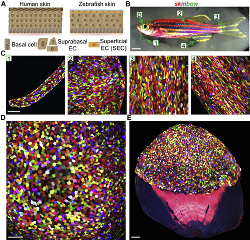
Multicolor Labeling of Superficial Epithelial Cells in skinbow Zebrafish (A) Schematic drawing of human and zebrafish skin. Zebrafish skin is a simple stratified epithelium without keratinized layers, with a surface covered by transparent mucus. EC, epithelial cell. (B) Brightfield view of an adult skinbow animal. Green boxes indicate areas where the z stacked confocal images shown in (C) (D) were captured. Scale bars, 2 mm. (C) Multicolor labeling of skin epithelium in adult skinbow animals from areas shown in (B). Scale bars, 100 μm. (D and E) Multicolor labeling of corneal and scale epithelium in skinbow. A dissected scale is shown in (E). SECs cover the exposed portion of the scale (top). Scale bars, 100 μm. |
Reprinted from Developmental Cell, 36, Chen, C.H., Puliafito, A., Cox, B.D., Primo, L., Fang, Y., Di Talia, S., Poss, K.D., Multicolor Cell Barcoding Technology for Long-Term Surveillance of Epithelial Regeneration in Zebrafish, 668-80, Copyright (2016) with permission from Elsevier. Full text @ Dev. Cell