
Fig. S1
- ID
- ZDB-FIG-191022-8
- Publication
- Chen et al., 2016 - Multicolor Cell Barcoding Technology for Long-Term Surveillance of Epithelial Regeneration in Zebrafish
- Other Figures
- All Figure Page
- Back to All Figure Page
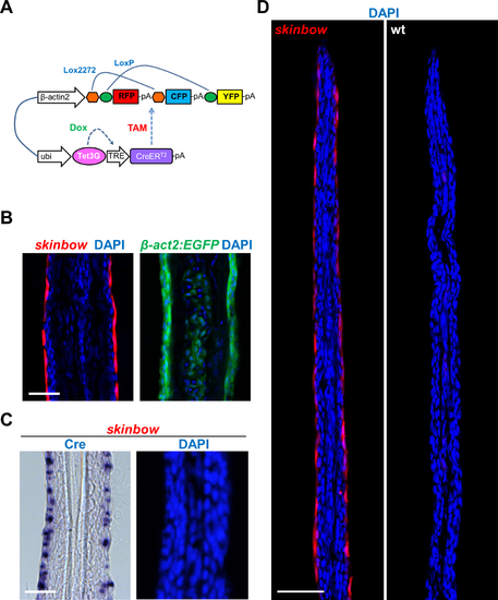
skinbow Labeling of the Outermost Layer of Adult Zebrafish Fin Epidermis, Related to Figure 1. (A) Single-transgene Brainbow-based construct. (B) Longitudinal sections of the caudal fin in skinbow and β- act2:EGFP lines. Although the Brainbow cassette is driven by the β-actin2 promoter in the skinbow construct, the expression is restricted to the SEC layer, likely a consequence of the transgene integration site. By contrast, EGFP fluorescence in the β-act2:EGFP line is present in all fin epithelial cell layers. (C) Longitudinal sections of skinbow fins assessed by in situ hybridization, visualizing CreER RNA expression in the SEC layers. (D) Longitudinal sections of distal tips of skinbow and wild-type caudal fins. Only the red channel is excited in these images. Scale bars, 50 μm. |
Reprinted from Developmental Cell, 36, Chen, C.H., Puliafito, A., Cox, B.D., Primo, L., Fang, Y., Di Talia, S., Poss, K.D., Multicolor Cell Barcoding Technology for Long-Term Surveillance of Epithelial Regeneration in Zebrafish, 668-80, Copyright (2016) with permission from Elsevier. Full text @ Dev. Cell