
|
Fig. 1
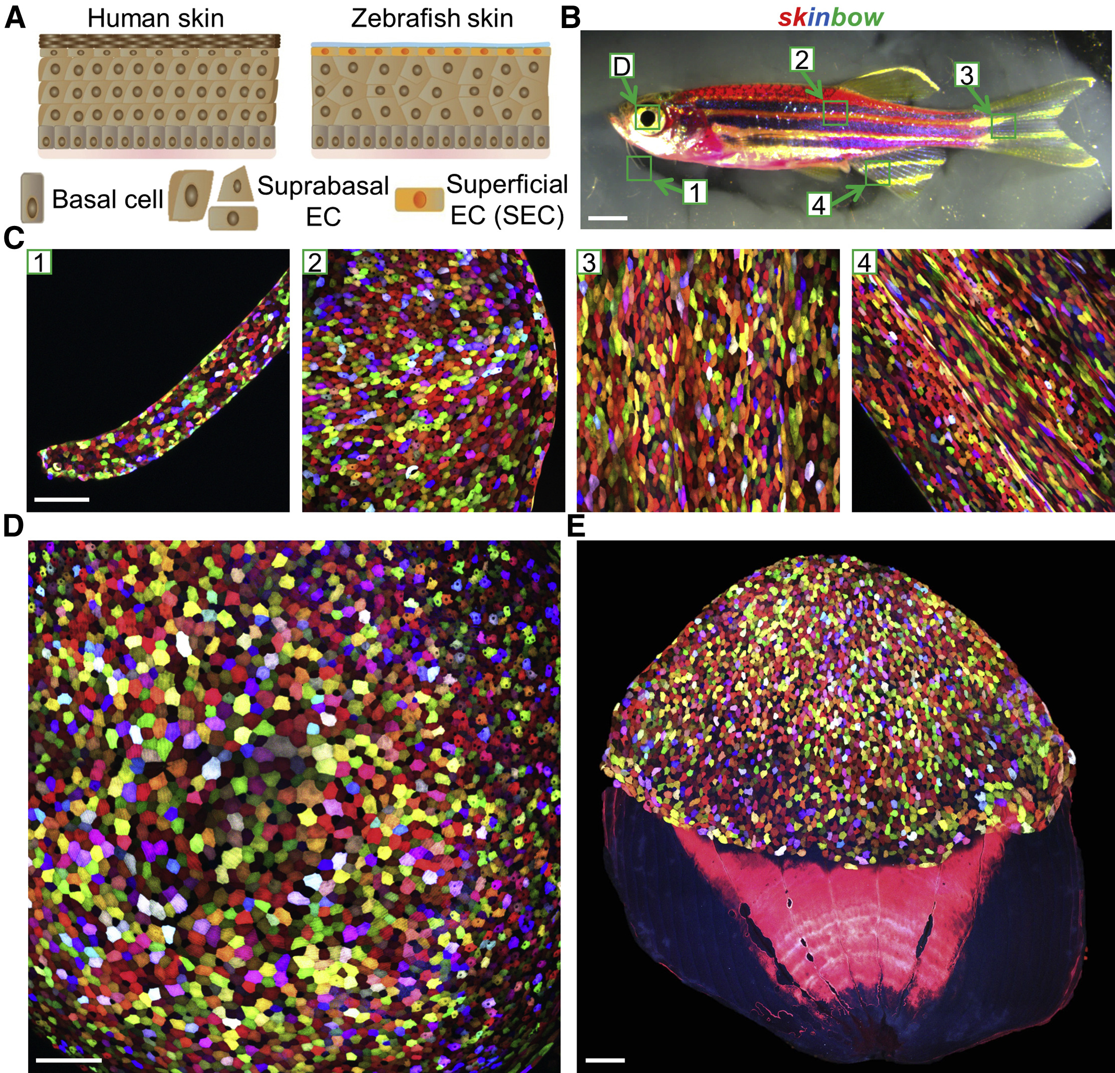
Multicolor Labeling of Superficial Epithelial Cells in skinbow Zebrafish
(A) Schematic drawing of human and zebrafish skin. Zebrafish skin is a simple stratified epithelium without keratinized layers, with a surface covered by transparent mucus. EC, epithelial cell.
(B) Brightfield view of an adult skinbow animal. Green boxes indicate areas where the z stacked confocal images shown in (C) (D) were captured. Scale bars, 2 mm.
(C) Multicolor labeling of skin epithelium in adult skinbow animals from areas shown in (B). Scale bars, 100 μm.
(D and E) Multicolor labeling of corneal and scale epithelium in skinbow. A dissected scale is shown in (E). SECs cover the exposed portion of the scale (top). Scale bars, 100 μm.
Reprinted from Developmental Cell, 36, Chen, C.H., Puliafito, A., Cox, B.D., Primo, L., Fang, Y., Di Talia, S., Poss, K.D., Multicolor Cell Barcoding Technology for Long-Term Surveillance of Epithelial Regeneration in Zebrafish, 668-80, Copyright (2016) with permission from Elsevier. Full text @ Dev. Cell