
Fig. S2
- ID
- ZDB-FIG-190812-37
- Publication
- Chen et al., 2019 - Cerebrovascular Injuries Induce Lymphatic Invasion into Brain Parenchyma to Guide Vascular Regeneration in Zebrafish
- Other Figures
- All Figure Page
- Back to All Figure Page
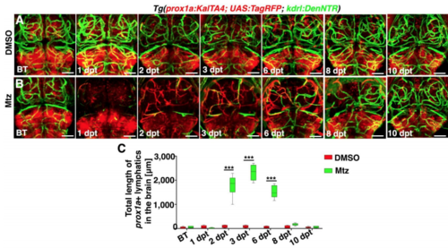
Time courses of meningeal lymphatic ingrowth and cerebrovascular regeneration shown by the prox1a promoter-driven transgene, Related to Figure 1 and Figure 2. (A–C) In contrast to DMSO treatment (A, n=35/36), a time course of meningeal lymphatic ingrowth and neoangiogenesis after Mtz treatment was exhibited (B, n=33/36). Images of 10 dpt showed a complete regeneration of brain vasculature. The quantifications show the total length of prox1a+ lymphatics (C, n=8. Two-way ANOVA by Dunnett’s multiple comparisons test. All P<0.0001). Dorsal view, anterior upward. Scale bar, 50 μm. Data are represented as mean ± SEM. ***P<0.001. |
Reprinted from Developmental Cell, 49(5), Chen, J., He, J., Ni, R., Yang, Q., Zhang, Y., Luo, L., Cerebrovascular Injuries Induce Lymphatic Invasion into Brain Parenchyma to Guide Vascular Regeneration in Zebrafish, 697-710.e5, Copyright (2019) with permission from Elsevier. Full text @ Dev. Cell