
Fig. 5
- ID
- ZDB-FIG-190812-31
- Publication
- Chen et al., 2019 - Cerebrovascular Injuries Induce Lymphatic Invasion into Brain Parenchyma to Guide Vascular Regeneration in Zebrafish
- Other Figures
- All Figure Page
- Back to All Figure Page
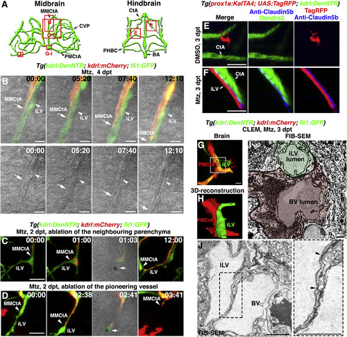
The Ingrown Lymphatics Act as “Growing Tracks” for Nascent Blood Vessels through Direct Physical Adhesion (A) Illustrations of midbrain and hindbrain vascular network indicate the image areas (red boxes) in panels. All panels are dorsal views, anterior upward. (B) Time-lapse imaging showed that the nascent blood vessel (kdrl+, MMCtA, arrows) grew along the preformed lymphatic vessel (fli1+kdrl−, arrowheads). Bright field indicated the two vessel structures. The elapsed time is indicated in h:min. Scale bar, 10 μm. See also Video S3. (C and D) Ablation of the ingrown lymphatic vessel (D) (n = 20/20 vessels in 10 larvae, 2 vessels per larvae) but not the neighboring parenchyma (C) (n = 20/20 sites in 10 larvae, 2 sites per larvae), led to the growth arrest and regression of the adhering nascent blood vessel. Arrowheads indicate the laser irradiation sites. The elapsed time is indicated in h:mins. Scale bar, 20 μm. See also Video S4. (E and F) Triple labeling of anti-Claudin5b antibodies, TagRFP and Dendra2 epifluorescence at 3 dpt. In contrast to DMSO treatment (E) (n = 21/22), Claudin5b was enriched at the interface between the ingrown lymphatic (prox1a+) and nascent blood vessels (kdrl+) after Mtz treatment (F) (n = 18/21). Scale bar, 20 μm. See also Figure S4. (G–J) Correlative light and electron microscopy (CLEM) of brain vessels at 6 dpf/3 dpt. The FIB-SEM target areas inside confocal images (G) are indicated with frames. Scale bar, 10 μm. 3D reconstructions of FIB-SEM single plans identify ingrown meningeal lymphatic vessel (mLV) and nascent blood vessel (BV) as two adhering vessel structures (H) (n = 3/3). The framed area in (G) is shown in (H). Single FIB-SEM image plane indicates the lumens of mLV and BV (I) (n = 3/3). Light green and light red mark the cross sections of the lymphatic vessel and blood vessel, respectively. The dotted frame in (J) indicates the border area of lymphatic and blood vessels, which is enlarged to show tight junctions (J) (arrows). Scale bar, 1 μm. See also Video S5. BA, basilar artery; CtA, central artery; CVP, choroidal vascular plexus; iLV, ingrown lymphatic vessels; MMCtA, middle mesencephalic central artery; PHBC, primordial hindbrain channel; PMCtA, posterior (caudal) mesencephalic central artery. |
Reprinted from Developmental Cell, 49(5), Chen, J., He, J., Ni, R., Yang, Q., Zhang, Y., Luo, L., Cerebrovascular Injuries Induce Lymphatic Invasion into Brain Parenchyma to Guide Vascular Regeneration in Zebrafish, 697-710.e5, Copyright (2019) with permission from Elsevier. Full text @ Dev. Cell