|
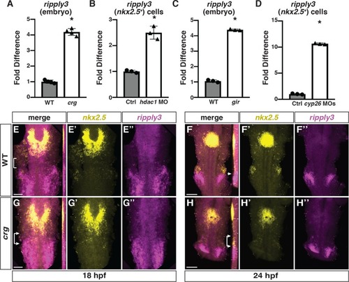
<italic>Ripply3</italic> is expanded anteriorly into <italic>nkx2</italic>.<italic>5+</italic> cells in <italic>crg</italic> mutants.(A-D) RT-qPCR for ripply3 expression in whole embryos at 48 hpf and sorted nkx2.5:ZsYellow+ cells at 28 hpf. (E-H”) Confocal images of two-color FISH for nkx2.5 and ripply3 in WT and crg mutant embryos at 18 and 24 hpf. Images are dorsal views with anterior up. Insets in F-H indicate lateral views of the confocal images. Bracket in E indicates space between posterior nkx2.5 and anterior ripply3 domains. Arrow in F indicates border between nkx2.5 in pharyngeal mesoderm and ripply3 in pharyngeal endoderm. Brackets with arrows in G and H indicate overlap in nkx2.5 and ripply3 domains in crg mutant embryos. n = 22 WT and n = 5 crg mutants embryos for 18 hpf and n = 19 WT and n = 11 crg mutants embryos for 24 hpf examined. Scale bars in E and G are 50 μm. Scale bars in F and H are 100 μm.
|