ZFIN ID: ZDB-FIG-190716-21 |
Antibody: | |
---|---|
Fish: | |
Condition: | |
Anatomical Term: | |
Stage: | Days 7-13 |
Fish: | |
---|---|
Condition: | |
Observed In: | |
Stage: | Days 7-13 |
Fig. 5 
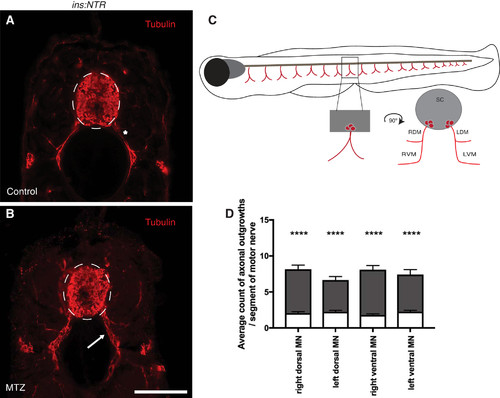
Peripheral motor axons appear defasciculated in hyperglycemic larvae. A and B: following the schedule of treatment described in Fig. 1A, axons (red, acetylated α-tubulin) appear defasciculated in the ins:NTR metronidazole (MTZ)-treated group (B, arrow) compared with the ins:NTR control group (A, asterisk). Dashed lines outline the spinal cord. NTR, nitroreductase. Scale bar = 50 μm. C: schematic of the areas in which axonal outgrowths were quantified: left dorsal muscle (LDM), right dorsal muscle (RDM), left ventral muscle (LVM), right ventral muscle (RVM), and spinal cord (sc). D: no differences were observed between segments within the groups. Number of axonal outgrowths was significantly greater in MTZ-treated (gray bars) than control (white bars) segments. MN, motor nerve. ****P < 0.0001. |
Antibody | Assay | Fish | Conditions | Stage | Anatomy |
---|---|---|---|---|---|
Ab1-tuba | IHC | jh4Tg | control | Days 7-13 | motor neuron axon |
IHC | jh4Tg | chemical ablation: pancreatic B cell, chemical treatment by environment: metronidazole | Days 7-13 | motor neuron axon |
Fish | Conditions | Stage | Phenotype |
---|---|---|---|
jh4Tg | chemical ablation: pancreatic B cell, chemical treatment by environment: metronidazole | Days 7-13 | motor neuron axon defasciculated, abnormal |
Acknowledgments: | This image is the copyrighted work of the attributed author or publisher, and ZFIN has permission only to display this image to its users. Additional permissions should be obtained from the applicable author or publisher of the image. Full text @ Am. J. Physiol. Regul. Integr. Comp. Physiol. |
---|