
FIGURE
Fig. 5
- ID
- ZDB-FIG-190702-26
- Publication
- Umali et al., 2019 - Loss of foxc1 in zebrafish reduces optic nerve size and cell number in the ganglion cell layer
- Other Figures
- All Figure Page
- Back to All Figure Page
Fig. 5
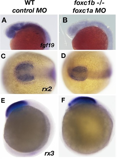
Expression of fgf19 is disrupted due to loss of foxc1function. fgf19 expression is found ubiquitously in the head at 20 hpf (A), and is reduced in foxc1b −/− injected with foxc1amorpholinos (B). The expression of two early retinal homeoboxgenes (rx2 and rx3) is not altered in these embryos (rx2, 16 somites C, D, rx3, 10 somites, E, F). Lateral views (A, B, E, F) and dorsal views (C, D). |
Expression Data
| Genes: | |
|---|---|
| Fish: | |
| Knockdown Reagent: | |
| Anatomical Terms: | |
| Stage Range: | 10-13 somites to 20-25 somites |
Expression Detail
Antibody Labeling
Phenotype Data
| Fish: | |
|---|---|
| Knockdown Reagent: | |
| Observed In: | |
| Stage: | 20-25 somites |
Phenotype Detail
Acknowledgments
This image is the copyrighted work of the attributed author or publisher, and
ZFIN has permission only to display this image to its users.
Additional permissions should be obtained from the applicable author or publisher of the image.
Reprinted from Vision Research, 156, Umali, J., Hawkey-Noble, A., French, C.R., Loss of foxc1 in zebrafish reduces optic nerve size and cell number in the ganglion cell layer, 66-72, Copyright (2019) with permission from Elsevier. Full text @ Vision Res.