
Fig. 1
- ID
- ZDB-FIG-190702-24
- Publication
- Umali et al., 2019 - Loss of foxc1 in zebrafish reduces optic nerve size and cell number in the ganglion cell layer
- Other Figures
- All Figure Page
- Back to All Figure Page
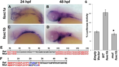
Zebrafish foxc1 genes. foxc1a is expressed in the neural crest and periocular mesenchyme at 24 hpf (A) and in the pharyngeal arches at 48 hpf (B). foxc1b is also expressed in the periocular mesenchyme and a small number of ocular progenitor cells at 24 hpf (C), and in the pharyngeal arches at 48 hpf (D). Partial alignment of the reference foxc1b coding sequence and the 40 base-pair deletion mutant sequence created by CRISPR genome editing (E). The zebrafish mutant strain deleted 40 nucleotides (57–96 of the open reading frame), resulting in a frameshift mutation and predicted premature protein truncation (F). Luciferase assays in HEK293 cells using a reporter with 6 copies of the consensus FOXC1 binding element demonstrates the zebrafish foxc1b 40 base pair deletion creates a null mutation, as no luciferase activity (above endogenous levels) is observed (G) p = 9 x 10⁻⁵. |
| Genes: | |
|---|---|
| Fish: | |
| Anatomical Terms: | |
| Stage Range: | Prim-5 to Long-pec |
Reprinted from Vision Research, 156, Umali, J., Hawkey-Noble, A., French, C.R., Loss of foxc1 in zebrafish reduces optic nerve size and cell number in the ganglion cell layer, 66-72, Copyright (2019) with permission from Elsevier. Full text @ Vision Res.