
Fig. 2
- ID
- ZDB-FIG-190624-8
- Publication
- Cao et al., 2019 - Germline Stem Cells Drive Ovary Regeneration in Zebrafish
- Other Figures
- All Figure Page
- Back to All Figure Page
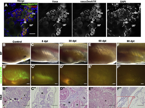
Ovary Regeneration Is Defective after Genetic Ablation of GSCs and Sex Reversal Finally Occurs (A) Comparison of Vasa protein and vasa:DenNTRexpression in ovary shows the Dendra2 fully overlaps with Vasa, suggesting that vasa:DenNTR is able to label all of the germ cells. (B–B″) The Tg(vasa:DenNTR)cq41 females of about 3 months old have big ovaries in bright field (BF) (B), strong green fluorescent (B′), and different stage germ cells (B″). (C–C″) At 5 dpt, the ovarian size largely decreases (C and C′) and except stage I and II oocytes, the germ cells are ablated by Mtz (C″). (D–D″) The ovaries become large and have mature oocytes at 30 dpt (D) but little green fluorescent is observed (D′), and the early stage germ cells were not detected (D″). (E–E″) By 60 dpt, the fish ovaries only have stage V cells. (F–F″) Testis (arrowheads) that lack germ cells appears at 90 dpt, showing that the females treated with Mtz reverted to sterile males. Oo, oogonia; IAz, zygotene-stage-IA; IA, stage IA oocytes; IB, stage IB oocyte; II, stage II oocyte; III, stage III oocyte; IV, stage IV oocyte; V, stage V oocyte. Scale bars: 50 μm (A), 1 mm (B–F′), 200 μm (B″–F″). See also Figure S2. |
| Fish: | |
|---|---|
| Condition: | |
| Observed In: | |
| Stage: | Adult |