
Fig. S10
- ID
- ZDB-FIG-190108-30
- Publication
- Abdul-Wajid et al., 2018 - Loss of embryonic neural crest derived cardiomyocytes causes adult onset hypertrophic cardiomyopathy in zebrafish
- Other Figures
- All Figure Page
- Back to All Figure Page
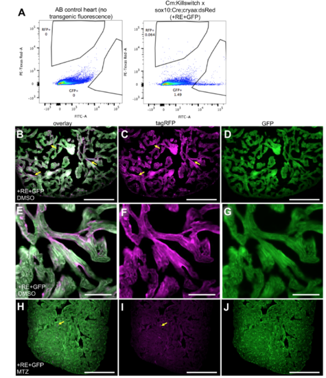
NC-Cm labeling in adult transgenic fish. A) Flow cytometry analysis and quantification of labeled cardiomyocytes in Cm:KillSwitch x Tg(sox10:cre;cryaa:dsRed) individual 15 adult heart versus AB control. GFP and RFP gates were drawn based on non-transgenic/non-fluorescent AB wild-type hearts. Dissociated cells were gated on viability (DAPI) and singlets before analyzing GFP and RFP populations. Three or more individual hearts were similarly analyzed by flow cytometry to quantify numbers of GFP+ and RFP+ cardiomyocytes. GFP+ and RFP+ percentages were added to quantify total number of cardiomyocytes in a whole heart and then RFP+ numbers were divided by this to generate their percent contribution to the total adult cardiomyocyte population. The average values of these quantifications are listed in Figure 4A and C. Heart dissociation protocol was carried out as previously described2. B-D) Adult sections of ventricles from control (+RE+GFP, DMSO). Sections were stained with anti-tagRFP and anti-GFP antibody. Fluorescent images were captured at 20X magnification. Yellow arrows point to tagRFP+ NC-Cms in trabeculae of adult heart. E-G) Higher magnification of tagRFP+ trabeculae from control hearts as in B-D. Scale bar =50uM H-J) NC-Cm ablated (+RE+GFP, MTZ) heart sections and example of remnant tagRFP (yellow arrow) seen in the NC-Cm ablated ventricle sections. Scale Bar = 200uM. |