
Fig. S3
- ID
- ZDB-FIG-190108-26
- Publication
- Abdul-Wajid et al., 2018 - Loss of embryonic neural crest derived cardiomyocytes causes adult onset hypertrophic cardiomyopathy in zebrafish
- Other Figures
- All Figure Page
- Back to All Figure Page
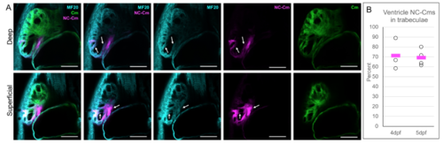
A) Cardiomyocyte labelling of Tg(Cm:KillSwitch) crossed to Tg(sox10:Cre;cryaa:dsRed). 5dpf embryos were immunostained with anti-sarcomere myosin heavy chain antibody MF20 and imaged in conjunction with tagRFP and GFP fluorescence. Confocal slices of deep (top panel) and superficial (bottom panel) areas of the heart are shown. Arrows point to examples of MF20 stain overlap with the NC-Cm tagRFP fluorescence. Scale bar = 50uM. B) Quantification of the percent of NC-Cms found in trabeculae of the ventricle relative to total ventricle NC-Cms. tagRFP+ NC-Cms were counted from 4 and 5dpf ventricles and designated as being within a trabeculae or not. The number found within trabeculae was divided by the total NC-Cms in the ventricle to yield percentage contribution. Circles represent individual hearts that were counted and bars represent mean of individual data points. |