
Fig. 5
- ID
- ZDB-FIG-190107-17
- Publication
- Crespo et al., 2018 - Characterisation of maturation of photoreceptor cell subtypes during zebrafish retinal development
- Other Figures
- All Figure Page
- Back to All Figure Page
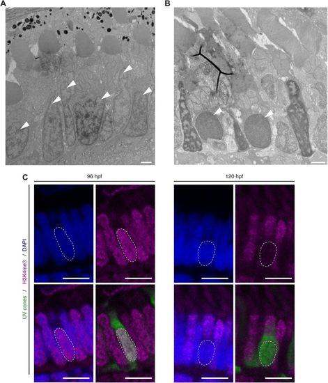
UV sensitive cones show changes in chromatic organisation. (A,B) Electron micrographs of sections through the PRC layer in the central retina of a wild-type zebrafish embryos at 96 hpf (A) and 120 hpf (B). Nuclei of some PRCs show alteration in chromatin at 120 hpf (arrowheads). (C) Confocal images of the PRC layer in retinal sections of Tg(-5.5opn1sw1:EGFP) embryos at 96 hpf and 120 hpf, with DAPI in blue, antibody staining for H3K4me3 in magenta and endogenous GFP fluorescence in green. UVS positive cells (green) show no staining for H3K4me3, a marker for euchromatin. Dashed lines mark the nucleus outline of a UVS cone. Scale bars: A–B: 1 µm, C: 5 µm. |