
Fig. S4
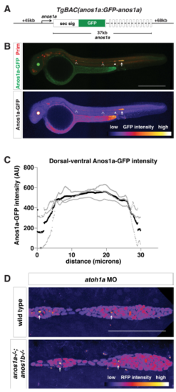
Characterization of anos1a:anos1a-GFP transgenic line Related to Figure 4. (A) Schematic of the anos1a BAC transgene. GFP was placed behind the Anos1a secretion signal in exon 1 of the anos1a locus. Numbers to the left and right indicate length of BAC upstream of exon 1 and downstream of exon 14 (the last exon), and number below indicates the distance between exon 1 and 14. White rectangles represent exons, and solid black lines in-between represent introns. (B) Top, immunostaining against GFP (green) and mCherry (red) of anos1a:GFPanos1a; prim:lyn2mCherry embryos. Bottom, false-coloring of immunostaining against Anos1a-GFP with intensity scale below. YFP expression in the lens originates from the transgenesis marker cryaa:citrine on the anos1a:anos1a-GFP transgene. Arrowheads indicate neuromasts with Anos1a-GFP signal, hollow arrowheads indicate neuromasts without Anos1a-GFP signal and the arrow indicates the primordium. Scale bar = 500 μm. (C) Anos1a-GFP intensity along the dorsal-ventral axis of the primordium. Gray lines represent individual embryos and the black line indicates the average. (D) Lumen integrity in embryos of genotypes indicated as assessed by accumulation of hsp70:sec-mCherry (arrows) in a primordium with nuclei marked by cxcr4b:H2AmCherry. Image is a maximum intensity projection of a Z-stack and is false colored based on fluorescence intensity using the fire look-up table (bottom). Sec-mCherry is distinguished from H2A-mCherry based on anatomical position, smaller size and higher signal intensity of microlumina compared to nuclei. Scale bar = 100 μm. |
| Genes: | |
|---|---|
| Fish: | |
| Anatomical Terms: | |
| Stage: | Prim-15 |
Reprinted from Developmental Cell, 46(6), Wang, J., Yin, Y., Lau, S., Sankaran, J., Rothenberg, E., Wohland, T., Meier-Schellersheim, M., Knaut, H., Anosmin1 Shuttles Fgf to Facilitate Its Diffusion, Increase Its Local Concentration, and Induce Sensory Organs, 751-766.e12, Copyright (2018) with permission from Elsevier. Full text @ Dev. Cell