
Fig. 4
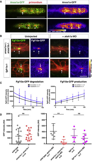
Anos1 Is Required for Luminal Accumulation of Fgf10a (A) Left, Anos1a-GFP protein (green) in the primordium marked with prim:lyn2mCherry (red). Right, Anos1a-GFP only in false colors. Top, arrow indicates Anos1a-GFP in microlumen. Bottom, arrowhead indicates Anos1a-GFP signal above apical constrictions. Scale bar represents 100 μm. (B) Live images of microluminal Fgf10a-GFP with membrane marker and false coloring of Fgf10a-GFP signal only in embryos of the indicated genotypes. Arrows indicate Fgf10a-GFP signal in microlumina and arrowheads (left) indicate Fgf10a-GFP-producing central cells adjacent to the microlumen. Central cells are missing in embryos injected with atoh1a morpholino (right). Scale bar represents 10 μm. (C) Total Fgf10a-GFP fluorescence intensity in the microlumen (left) and central cells (right) when secretion is blocked with brefeldin A. Error bars indicate the SD. Fgf10a-GFP degradation was fitted to a one-phase decay model with half-life values of 102 min (95% CI 33 to 197 min) for heat-shocked control embryos and 79 min (95% CI 25 to 157 min) for anos1b-over-expressing embryos. Fgf10a-GFP production was fitted to a linear model with a production rate of 75.2 ± 8.2 min−1 for heat-shocked control embryos and a production rate of 89.2 ± 11.8 min−1 for anos1b-over-expressing embryos. (D) Total Fgf10a-GFP intensity in mature microlumina at the fourth apical constriction from the front in uninjected embryos (left) and embryos injected with atoh1a morpholino (right). Mean, SD, and individual data points are shown. n.s. = p > 0.05, ∗∗ = p < 0.01. ANOVA p < 0.05. See also Figure S4 and Videos S4 and S5. |
| Genes: | |
|---|---|
| Fish: | |
| Knockdown Reagent: | |
| Anatomical Term: | |
| Stage: | Prim-15 |
| Fish: | |
|---|---|
| Knockdown Reagent: | |
| Observed In: | |
| Stage: | Prim-15 |
Reprinted from Developmental Cell, 46(6), Wang, J., Yin, Y., Lau, S., Sankaran, J., Rothenberg, E., Wohland, T., Meier-Schellersheim, M., Knaut, H., Anosmin1 Shuttles Fgf to Facilitate Its Diffusion, Increase Its Local Concentration, and Induce Sensory Organs, 751-766.e12, Copyright (2018) with permission from Elsevier. Full text @ Dev. Cell