
Fig. S6
- ID
- ZDB-FIG-181019-45
- Publication
- Yin et al., 2018 - Spatiotemporal Coordination of FGF and Shh Signaling Underlies the Specification of Myoblasts in the Zebrafish Embryo
- Other Figures
- All Figure Page
- Back to All Figure Page
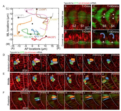
The apparent somite rotation is underpinned by the sequential cell shape changes and muscle elongation, related to Figure 5 and 7 (A) Cell tracking of 8 lateral somitic cells at the coronal plane of (B’) from stage S1 to 8 hours later after the completion of the primary myogenesis. The tracks are displayed with lateral to the top and anterior to the left. The initial positions of the cells are labelled with rectangles with specific colour to the track of each cell. Four relative medial cells are denoted as cell M1, M2, M3 and M4. The other four relative lateral cells are denoted as cell L1, L2, L3 and L4. The black circle denotes the centre of the somite. The angle of rotation of each individual cell relative to the somite centre is labelled in the figure. (B-C) parasagittal (B and C) and coronal (B’ and C’) planes of somite at stage of S2 and S1 (B and B’) and 90 mins later (C and C’). The coronal planes are taken at the positions of the dash lines in the parasagittal planes. Slow muscles are labelled with Prdm1a:GFP. White short arrows denote the posterior fast muscle progenitors that move medially. White dash lines label the contours of slow muscles in (B’ and C’). The blue arrows indicate the direction of the initial apparent somite rotation, prior to slow muscle migration. (D-F) Cell tracking of neighboring lateral somitic cells at distinct locations. (D) Cells located at the lateral surface of the somite. (E) Fast muscle progenitors located at the middle of the somite. (F) Fast muscle progenitors located at the posterior of the somite. White arrow in E (iii) denotes a cell division. White asterisks in F(ii) and F(iii) denote the somitic cells moving medially from the lateral somite. |
Reprinted from Developmental Cell, 46, Yin, J., Lee, R., Ono, Y., Ingham, P.W., Saunders, T.E., Spatiotemporal Coordination of FGF and Shh Signaling Underlies the Specification of Myoblasts in the Zebrafish Embryo, 735-750.e4, Copyright (2018) with permission from Elsevier. Full text @ Dev. Cell