
Fig. 4
- ID
- ZDB-FIG-181019-36
- Publication
- Yin et al., 2018 - Spatiotemporal Coordination of FGF and Shh Signaling Underlies the Specification of Myoblasts in the Zebrafish Embryo
- Other Figures
- All Figure Page
- Back to All Figure Page
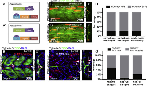
The Direct and Indirect Roles of FGF Signaling in the Further Differentiation of Fast and Slow Lineages, Respectively (A–D) Mosaic FGF perturbations in slow muscle cells driven by smyhc1:gal4;UAS:dn-fgfr1-p2a-mCherry (A) or smyhc1:gal4;UAS:ca-fgfr1-p2a-mCherry (Aʹ), respectively. (B and C) Mosaic FGF inhibition (B) or over-activation (C) in the slow muscle fibers is identifiable through mCherry expression. Images are taken at 28 hpf at muscle segments 13–16 and projected along ML axis. (Bʹ–Cʹ) Transverse views of (B and C) made at the sites of dash lines with lateral to the left and dorsal to the top. (D) Fraction of mCherry positive MPs among mCherry positive slow muscles in control group (60/296), FGF inhibited group (52/262), and FGF over-activated group (45/214). (E–G) Mosaic FGF perturbations regardless of cell types with the expression of dn-fgfr1-mCherry (E) or ca-fgfr1-p2a-mCherrry (F) driven by heat shockpromoter hsp70l. The perturbed cells are identifiable through membrane localized dn-fgfr1-mCherry (E) or uniformly distributed mCherry, respectively (F), and labeled with white short arrows. (Eʹ–Fʹ) Transverse views of (E and F) made at the sites of dash lines with lateral to the left and dorsal to the top. (G) Fraction of mCherry positiveECL cells among the total population of mCherry positive cells in fast muscle lineage (both fast muscle fibers and ECL cells) in control group (104/495), FGF inhibited group (373/777), and FGF over-activated group (41/696). Heat shock was performed at 16-somite stage. The quantification of cell fates was performed at muscle segments 16–22 at 30 hpf. |
Reprinted from Developmental Cell, 46, Yin, J., Lee, R., Ono, Y., Ingham, P.W., Saunders, T.E., Spatiotemporal Coordination of FGF and Shh Signaling Underlies the Specification of Myoblasts in the Zebrafish Embryo, 735-750.e4, Copyright (2018) with permission from Elsevier. Full text @ Dev. Cell