
Fig. 3
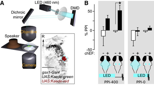
Optogenetic Activation of Gsx1 Neurons in the PPI-Active Zone Inhibits Startle Responses (A) Schematic of experimental setup for selective illumination during behavioral prepulse inhibition. Inset: Kaede photoconversion in Gsx1 neurons (gsx1-Gal4, UAS:Kaede) by targeted illumination (cyan diamond). Scale bar, 50 μm. (B) Prepulse inhibition after LED illumination of the hindbrain (large square, cyan), or R4 alone (small squares, cyan) in larvae with Gsx1 cells expressing chEF (black), and clutchmates lacking chEF (white). Left panel: LED illumination terminating 400 ms before the acoustic startle stimulus (PPI-400, n = 7 chEF+, n = 3 chEF–). Main effect of chEF expression using repeated-measures ANOVA on rank-transformed %PPI F1,7 = 10.8, p = 0.013. Error bars are SEM. Right panel: LED illumination immediately before and during an acoustic pulse (PPI-0, n = 5 chEF+, n = 3 chEF–). Main effect of chEF expression F1,6 = 3.3, p = 0.12. ∗p < 0.05 Mann-Whitney test. Error bars are SEM. See also Figure S2 and Table S1. |