
Fig. 7
- ID
- ZDB-FIG-180720-37
- Publication
- Löhr et al., 2018 - Diet-Induced Growth Is Regulated via Acquired Leptin Resistance and Engages a Pomc-Somatostatin-Growth Hormone Circuit
- Other Figures
- All Figure Page
- Back to All Figure Page
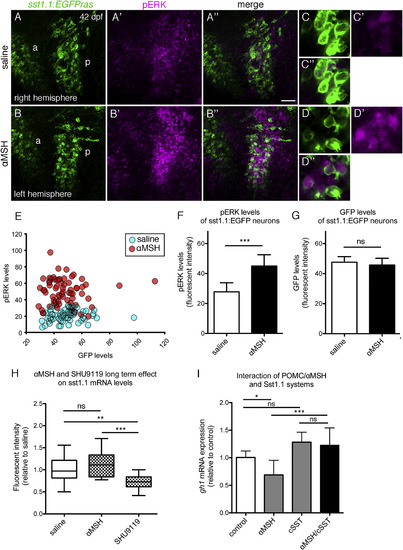
Zebrafish Sst Neurons Respond to αMSH to Mediate αMSH’s Effect on GH Expression (A–G) pERK response of hypophysiotropic Sst1.1 cells to αMSH treatment in sst1.1:EGFPras fish (42 dpf). (A–B′′) POA of two hemispheres derived from the same brain, differentially incubated in saline (A–A′′) or 10 μM αMSH (B–B′′) for 30 min. Double IF for GFP (green) and pERK (magenta) reveals increase in pERK levels after αMSH application in the posterior POA (p). Images of the right hemisphere were flipped horizontally. (C–D′′) Magnification of posterior POA Sst1.1 neurons without (C–C′′) or with (D–D′′) αMSH treatment. (E–G) Quantification of pERK (E and F) and GFP (E and G) levels in a total of seven brain hemisphere pairs as shown in (A) and (B), revealing significantly increased pERK, but unaltered GFP levels in posterior Sst1.1 neurons after αMSH treatment. (H) Effect of long-term αMSH or SHU9119 treatment on sst1.1 mRNA levels. sst1.1 FISH and signal intensity determination from confocal images of juvenile fish (42 dpf) that had been subjected to three repetitive ICV injections of αMSH, SHU9119, or saline over the course of 48 hr. (I) Epistasis between Pomca/αMSH and Sst systems; gh1 expression levels assessed by qRT-PCR analysis after treatment of larvae (14 dpf) for 8 hr with 10 μM αMSH, 1 μM cSST, or a combination of both peptides. Scale bars represent 50 μm (A–B′′) and 10 μm (C–D′′). ∗p < 0.05; ∗∗p < 0.01; ∗∗∗p < 0.001. Error bars in (F)–(I) show SD. |
| Gene: | |
|---|---|
| Antibody: | |
| Fish: | |
| Condition: | |
| Anatomical Term: | |
| Stage: | Days 30-44 |
| Fish: | |
|---|---|
| Condition: | |
| Observed In: | |
| Stage: | Days 30-44 |