
FIGURE
Fig. S2
- ID
- ZDB-FIG-180711-57
- Publication
- Iwasaki et al., 2018 - Epidermal regulation of bone morphogenesis through the development and regeneration of osteoblasts in the zebrafish scale
- Other Figures
- All Figure Page
- Back to All Figure Page
Fig. S2
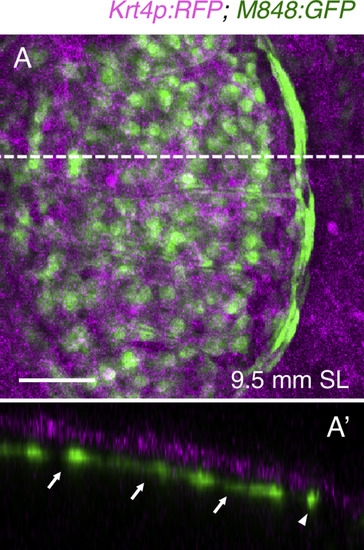
M848:GFP is expressed in the dermis. (A) Expression of GFP and RFP in juvenile krt4:RFP;M848:GFP fish, with the epidermis labeled by RFP. (A′) Optical cross-section (at a thickness of 76.2 µm) along the dashed line in (A). Both central cells (arrows) and marginal cells (arrowhead) are located in the dermis. Scale bar, 50 µm. |
Expression Data
Expression Detail
Antibody Labeling
Phenotype Data
Phenotype Detail
Acknowledgments
This image is the copyrighted work of the attributed author or publisher, and
ZFIN has permission only to display this image to its users.
Additional permissions should be obtained from the applicable author or publisher of the image.
Reprinted from Developmental Biology, 437(2), Iwasaki, M., Kuroda, J., Kawakami, K., Wada, H., Epidermal regulation of bone morphogenesis through the development and regeneration of osteoblasts in the zebrafish scale, 105-119, Copyright (2018) with permission from Elsevier. Full text @ Dev. Biol.