|
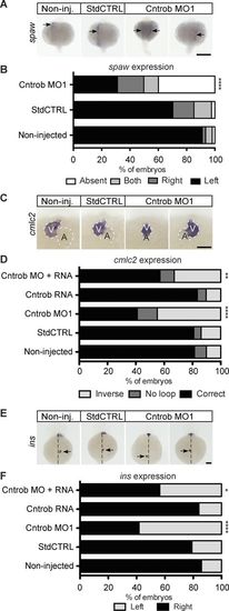
Centrobin loss causes laterality defects in zebrafish embryos. (A, C, and E) WMISH micrographs of showing localization of the messages of spaw for left lateral plate mesoderm, cmlc2 to indicate heart looping, and ins to label pancreata and assess abdominal situs development. Dashed line indicates midline. A, atrium; V, ventricle. Bars, 100 µm. (B) Quantitation of laterality of spaw expression (arrows) in WT embryos and embryos injected as indicated at 20–22 ss. Appropriate asymmetric expression is to the left. (D) Quantitation of appropriate heart looping in WT embryos and embryos injected as indicated at 48 hpf. (F) Quantitation of pancreas placement in WT embryos and embryos injected as indicated at 48 hpf. Correct placement is on the right side from the midline. *, P < 0.05; **, P < 0.01; ****, P < 0.0001. Significances were assessed using Fisher’s exact test.
|