|
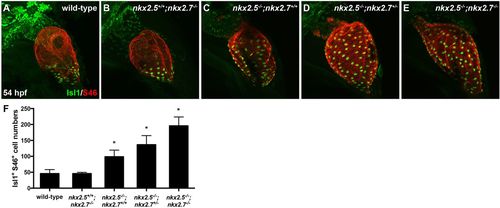
Nkx genes inhibit Isl1 expression at the sinus venosus. (A-E) Lateral view, anterior towards the top, at 54 hpf in wild-type (A), nkx2.5+/+;nkx2.7−/− (B), nkx2.5−/−;nkx2.7+/+ (C), nkx2.5−/−;nkx2.7+/− (D) and nkx2.5−/−;nkx2.7−/− (E) embryos. Representative confocal images of immunofluorescence for Isl1 (green) and S46 (red) demonstrate that Isl1 is restricted to the venous pole of wild-type (A) and nkx2.5+/+;nkx2.7−/− (B) embryos. However, Isl1 expression expands throughout the atrium in nkx mutants (C-E). (F) Quantification of Isl1+ S46+ CMs is depicted showing a gradual increase in cell number with a progressive loss of nkx alleles. Mean and s.e.m. of each data set are shown (*P<0.005) in wild-type (n=16), nkx2.5+/+;nkx2.7−/− (n=2), nkx2.5−/−;nkx2.7+/+ (n=6), nkx2.5−/−;nkx2.7+/− (n=10) and nkx2.5−/−;nkx2.7−/− (n=4) embryos. Student's t-test was used to determine statistical significance.
|