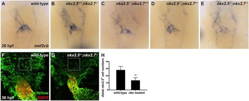
Nkx genes delimit the OFT progenitor pool. (A-E) Dorsal view, anterior towards the top, at 26 hpf. In situ hybridization depicts diminished expression of mef2cb, which labels aSHF-derived CM progenitors, in representative nkx2.5−/−;nkx2.7+/+ (n=6) (C), nkx2.5−/−;nkx2.7+/− (n=3) (D) and nkx2.5−/−;nkx2.7−/− (n=5) (E) embryos compared with wild-type (n=15) (A) and nkx2.5+/+;nkx2.7−/− (n=5) (B) embryos. (F,G) Ventral view of the arterial pole region at 36 hpf. Confocal projections of immunohistochemistry for ZsYellow (green) and EGFP (red) in embryos carrying Tg(nkx2.5:ZsYellow) and Tg(myl7:EGFP) illustrate decreased contribution of nkx2.5+ aSHF-derived CM progenitors (ZsYellow+ EGFP−) to the arterial pole in nkx2.5−/−;nkx2.7−/− (G) compared with wild-type (F) embryos (boxed areas). (H) Quantification of ZsYellow+ EGFP− cells at the arterial pole in wild-type (n=9) and nkx mutant (n=8) embryos reveals a statistically significant decrease in this population following the loss of Nkx gene function. Student's t-test was used to determine statistical significance. Mean and s.e.m. of each data set are shown (**P<0.0001).
|
