
Fig. 1
- ID
- ZDB-FIG-180516-12
- Publication
- Ojeda Naharros et al., 2017 - Loss-of-function of the ciliopathy protein Cc2d2a disorganizes the vesicle fusion machinery at the periciliary membrane and indirectly affects Rab8-trafficking in zebrafish photoreceptors
- Other Figures
- All Figure Page
- Back to All Figure Page
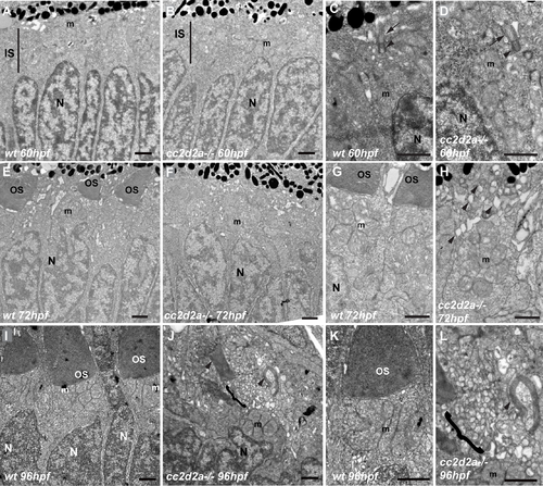
Vesicle accumulation is progressive from the onset of OS formation in cc2d2a-/- PRs (A-D) Transmission electron microscopy images (TEM) of retinal sections at 60 hpf are indistinguishable between wild-type (wt) (A, C) and cc2d2a-/- (B, D), including with respect to basal body docking (arrowheads in C-D) and extension of the connecting cilium (arrows in C-D). (E-H) Retinal sections at 72 hpf: note the nascent outer segments (OS) in wt (E, G), but the quasi-absence of OSs in cc2d2a-/- with onset of apical accumulation of vesicular structures (F, H, arrowheads). (I-L) Retinal sections at 96 hpf: well-formed OSs are present apical to the mitochondrial cluster in wt (I, K), while in mutant photoreceptors an increased number of vesicles (brackets) are found in the apical portion of the cell together with misshapen membrane stacks (arrowheads) instead of OSs (J, L). Scale bars are 1 μm in all panels. m mitochondria, N nuclei, OS outer segments, wt wild-type, hpf hours post fertilization. |
| Fish: | |
|---|---|
| Observed In: | |
| Stage Range: | Protruding-mouth to Day 4 |