
Fig. 2
- ID
- ZDB-FIG-180501-2
- Publication
- Gross-Thebing et al., 2017 - The Vertebrate Protein Dead End Maintains Primordial Germ Cell Fate by Inhibiting Somatic Differentiation
- Other Figures
- All Figure Page
- Back to All Figure Page
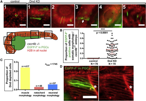
Loss of Dnd Function Results in Morphological Transdifferentiation (A) Dnd-deficient PGCs in 24-hpf cxcr4b−/− embryos exhibit different somatic morphologies based on the tissue within which they reside. A cartoon presenting a 24-hpf embryo in which distinct regions are framed and numbered. Images were captured from the following regions: 1, muscle; 2, eye; 3, brain; 4, otic capsule; 5, notochord. The preferential expression of EGFP on the membrane of PGCs is achieved by injection of RNA encoding for EGFP-F′ that contains the 3′ UTR of nanos3 (Köprunner et al., 2001). The arrow in 3 points to axon-like projections within neural tissue. Scale bars, 30 μm. (B) The proportion of PGCs showing somatic morphology in Dnd-deficient, cxcr4b−/− embryos at 24 hpf. p Values were determined using the Mann-Whitney U test with α = 0.05. Data are presented as median ± IQR. (C) Graph presenting the proportion of cells exhibiting specific somatic morphologies among all Dnd-depleted cells examined (ntotal). (D) Dnd-deficient PGCs exhibit morphology similar to that of somatic cells in their environment. Cells are labeled with farnesylated fluorescent proteins to direct the label to the membrane (F′). Confocal image captured at 24 hpf showing Dnd-deficient PGCs (asterisk) located within the developing muscle tissue (see Figure S2E for examples from other tissues). Scale bar, 50 μm. N, number of embryos; n, number of cells; F′, farnesylated; KD, knockdown; hpf, hours post fertilization. See also Figure S2. |
| Fish: | |
|---|---|
| Knockdown Reagents: | |
| Observed In: | |
| Stage: | Prim-5 |
Reprinted from Developmental Cell, 43, Gross-Thebing, T., Yigit, S., Pfeiffer, J., Reichman-Fried, M., Bandemer, J., Ruckert, C., Rathmer, C., Goudarzi, M., Stehling, M., Tarbashevich, K., Seggewiss, J., Raz, E., The Vertebrate Protein Dead End Maintains Primordial Germ Cell Fate by Inhibiting Somatic Differentiation, 704-715.e5, Copyright (2017) with permission from Elsevier. Full text @ Dev. Cell