
Fig. S1
- ID
- ZDB-FIG-180501-8
- Publication
- Gross-Thebing et al., 2017 - The Vertebrate Protein Dead End Maintains Primordial Germ Cell Fate by Inhibiting Somatic Differentiation
- Other Figures
- All Figure Page
- Back to All Figure Page
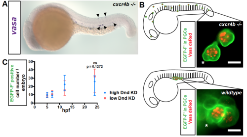
Related to Figure 1. Survival of PGCs at ectopic positions (A) Expression of vasa RNA in PGCs (blue) in cxcr4b-/- mutant embryos at 24 hpf by in situ hybridization. Arrowheads point at ectopic PGCs. (B) Characteristic bleb-like protrusions (asterisks) and germ cell granules (marked with Vasa-dsRed fusion protein, red) are observed in ectopic PGCs (membrane marked with EGFP-F’, green) in cxcr4b-/- embryos (upper panel) and in PGCs localized to the region of the gonad in wildtype embryos (lower panel). Scale bar, 10 μm. Images were captured at 19 hpf. See also movie S1. (C) A graph presenting the number of PGCs detected by expression of EGFP at different time points during the first day of development in embryos injected with either low (20 μM) or high (60 μM) amounts of dnd Morpholino. 50≤N≤61, Number of embryos (N). Data are presented as median ± IQR. Farnesylated (F’), Knockdown (KD), hours post fertilization (hpf). |
Reprinted from Developmental Cell, 43, Gross-Thebing, T., Yigit, S., Pfeiffer, J., Reichman-Fried, M., Bandemer, J., Ruckert, C., Rathmer, C., Goudarzi, M., Stehling, M., Tarbashevich, K., Seggewiss, J., Raz, E., The Vertebrate Protein Dead End Maintains Primordial Germ Cell Fate by Inhibiting Somatic Differentiation, 704-715.e5, Copyright (2017) with permission from Elsevier. Full text @ Dev. Cell