
Fig. S4
- ID
- ZDB-FIG-180424-6
- Publication
- Balasanyan et al., 2017 - Structure and Function of an Actin-Based Filter in the Proximal Axon
- Other Figures
- All Figure Page
- Back to All Figure Page
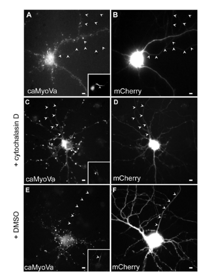
caMyoVa localization in the presence and absence of Cytochalasin D, related to Fig. 3. (A) Cortical neuron expressing caMyoVa, which is present mainly in the somatodendritic compartment in a diffuse manner (n = 10 neurons, 3 cultures). (B) Cortical neuron in (A) expressing mCherry. (C) Cortical neuron in culture expresses caMyoVa in the axon in the presence of Cytochalasin D. (n = 11 neurons, 3 cultures). (D) mCherry expressed in the same neuron as in (C). (E) caMyoVa is absent from the axon in the presence of DMSO. (n = 9 neurons, 3 cultures). (F) mCherry expressed in the same neuron as in (E). Insets show Ankyrin G staining. Arrowheads indicate axon. Scale bar 5 μm. |