
Fig. S1
- ID
- ZDB-FIG-180424-4
- Publication
- Balasanyan et al., 2017 - Structure and Function of an Actin-Based Filter in the Proximal Axon
- Other Figures
- All Figure Page
- Back to All Figure Page
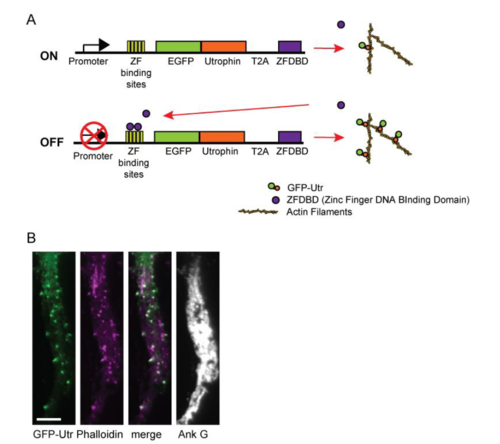
Regulated expression of Utrophin for labeling actin, related to Fig. 1. (A) Expression of Utrophin fused to GFP (GFP-Utr) and the CCR5 Zinc Finger DNA binding domain (ZFDBD) are driven by the CAG promoter. Just downstream of the transcriptional start site there are five DNA binding sites recognized by the CCR5 Zinc Finger (ZF binding site). Binding of the ZFDBD to the ZF binding site sterically hinders transcription of GFP-Utr limiting its expression. (B) Expression pattern of GFP-Utrophin in the proximal axon of a cortical neuron in culture (green) is highly similar to that of Phalloidin (purple), consistent with GFP-Utr faithfully labeling actin filaments. Ankyring G staining confirms that the process is an axon. Scale bar 5 μm. |