
|
Fig. S4
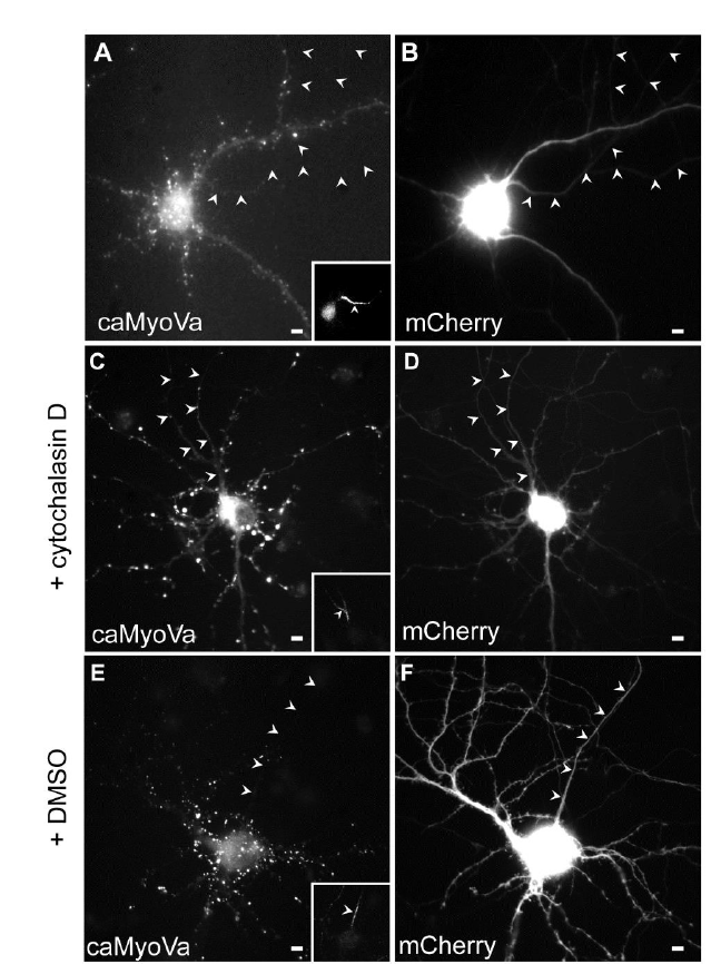
caMyoVa localization in the presence and absence of Cytochalasin D, related to Fig. 3. (A) Cortical neuron expressing caMyoVa, which is present mainly in the somatodendritic compartment in a diffuse manner (n = 10 neurons, 3 cultures). (B) Cortical neuron in (A) expressing mCherry. (C) Cortical neuron in culture expresses caMyoVa in the axon in the presence of Cytochalasin D. (n = 11 neurons, 3 cultures). (D) mCherry expressed in the same neuron as in (C). (E) caMyoVa is absent from the axon in the presence of DMSO. (n = 9 neurons, 3 cultures). (F) mCherry expressed in the same neuron as in (E). Insets show Ankyrin G staining. Arrowheads indicate axon. Scale bar 5 μm.