
Fig. 5
- ID
- ZDB-FIG-180404-30
- Publication
- Sakamaki et al., 2015 - Conservation of structure and function in vertebrate c-FLIP proteins despite rapid evolutionary change
- Other Figures
- All Figure Page
- Back to All Figure Page
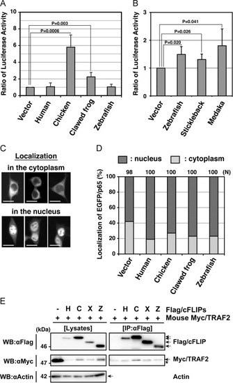
Functional analyses of non-mammalian c-FLIP proteins on NF-κB activation ability. (A, B) Enzymatic analysis of NF-κB activation induced by non-mammalian c-FLIP proteins. The empty vector or plasmids carrying c-FLIP were transiently co-transfected with pNFκB-Luc and pRL-TK into HEK293 cells, and cultured for 48 h. NF-κB activation was analyzed by measuring enzyme activities of dual luciferases produced in transfected cells using a luminometer. Data are presented as the means and standard deviations of samples counted from three independent experiments. The statistically-significant difference between two groups was evaluated by Student’s t-test. (C) Cytological analysis of a NF-κB component, p65, in cells expressing c-FLIP. HEK293 cells were transiently transfected with an empty vector or plasmids carrying c-FLIP together with pEGFP/p65, and cultured for 24 h. The localization of EGFP/p65 proteins in transfected cells was analyzed by fluorescence microscopy. Typical patterns of subcellular localization of EGFP/p65: EGFP/p65 normally localizes in the cytoplasm (upper panels), but it translocates into the nucleus when the NF-κB signaling pathway undergoes activation (lower panels) [35]. Scale bars indicate 20 μm. (D) A summary of cytological analyses on the translocation of EGFP/p65. Within each field, positive cells (dark gray) and negative cells (light gray) for EGFP/p65 translocation were counted under the fluorescent microscope and percentages of total were calculated. N indicates the total number of transfected cells examined in four independent experiments. (E) Co-immunoprecipitation and immunoblot analysis of physical interactions between non-mammalian c-FLIP proteins and mouse TRAF2. HEK293 cells were transfected with either pCMV-Flag/HsFLIP, pME18S-Flag/GgFLIP, pME18S-Flag/XlFLIP, pME18S-Flag/DrFLIP, or control vector in combination with pME18S-Myc/TRAF2. Forty-eight hours after transfection, cells were lysed and the c-FLIP complex was immunoprecipitated from whole cell lysates with an anti-Flag antibody. Samples were analyzed alongside aliquots of the cell lysates by immunoblotting with anti-Flag, anti-Myc, and anti-actin antibodies, respectively. An asterisk indicates immunoglobulin light-chain. Abbreviations: H, human; C, chicken; F, clawed frog; Z, zebrafish; IP, immunoprecipitation; WB, western immunoblotting. |