
Fig. 4
- ID
- ZDB-FIG-180404-29
- Publication
- Sakamaki et al., 2015 - Conservation of structure and function in vertebrate c-FLIP proteins despite rapid evolutionary change
- Other Figures
- All Figure Page
- Back to All Figure Page
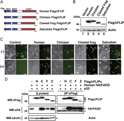
Assessment of the anti-apoptotic activity of the c-FLIP subfamily proteins. (A) The protein structure of Flag-tagged non-mammalian c-FLIP proteins consisting of two DED motifs and a CASc* protease-like domain. (B) Immunoblot analysis of non-mammalian c-FLIP proteins. The empty vector, pCMV-Flag/HsFLIP, pME18S-Flag/GgFLIP, pME18S-Flag/XlFLIP, or pME18S-Flag/DrFLIP were transiently transfected into HeLa cells. After culturing for 48 h, transgene products were analyzed by immunoblotting with an anti-Flag antibody. (C) Cytological analysis of transfectants expressing non-mammalian c-FLIP proteins. HeLa cells expressing Venus with or without human, chicken, clawed frog, and zebrafish c-FLIP were incubated in the presence (lower panels) or absence (upper panels) of anti-Fas antibody and CHX for 8 h, and examined for viability by monitoring Venus-positive cells. Both phase-contrast and fluorescence images in the same field were captured under the microscope. Scale bars represent 100 μm. (D) Co-immunoprecipitation and immunoblot analysis of physical interactions between non-mammalian c-FLIP proteins and human FADD. Human HEK293 cells were co-transfected with pME18S-HA/hFADD in conjunction with pME18S empty vector, pCMV-Flag/HsFLIP, pME18S-Flag/GgFLIP, pME18S-Flag/XlFLIP, or pME-Flag/DrFLIP. Baculovirus p35 was introduced into all transfected cells to prevent cell death. After 2 days of cultivation, transfected cells were harvested and lysed in lysis buffer. The cell lysates were immunoprecipitated with an anti-FLAG M2 affinity gel. Coimmunoprecipitates and aliquots of cell lysates were examined by immunoblot analysis with anti-Flag, anti-HA, and anti-actin antibodies, respectively. An asterisk indicates immunoglobulin light-chain. Abbreviations: H, human; C, chicken; F, clawed frog; Z, zebrafish; IP, immunoprecipitation; WB, western immunoblotting. |