
FIGURE
Fig. 4
- ID
- ZDB-FIG-180119-9
- Publication
- Chen et al., 2017 - Label-free photoacoustic imaging of the cardio-cerebrovascular development in the embryonic zebrafish
- Other Figures
- All Figure Page
- Back to All Figure Page
Fig. 4
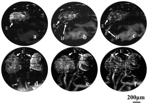
Vascular development in the heart and brain regions of the zebrafish embryo. (a-c): Longitudinal imaging of the heart in the embryonic zebrafish at 22, 25 and 28 hpf, respectively. (d-f): The sagittal view of the microvascular development in the brain at 36, 48 and 72 hpf, respectively. H: Heart E: Eye. |
Expression Data
Expression Detail
Antibody Labeling
Phenotype Data
Phenotype Detail
Acknowledgments
This image is the copyrighted work of the attributed author or publisher, and
ZFIN has permission only to display this image to its users.
Additional permissions should be obtained from the applicable author or publisher of the image.
Full text @ Biomed. Opt. Express