
- Title
-
Label-free photoacoustic imaging of the cardio-cerebrovascular development in the embryonic zebrafish
- Authors
- Chen, Q., Jin, T., Qi, W., Mo, X., Xi, L.
- Source
- Full text @ Biomed. Opt. Express
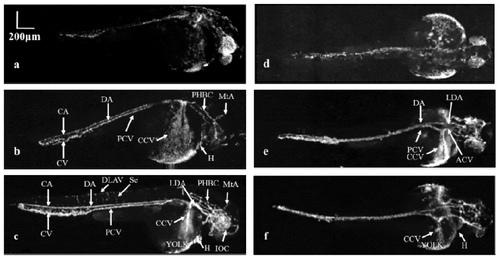
Vascular development of the zebrafish embryo from 36 to 72 hpf with sagittal (a-c) and coronal views (d-f) in two different zebrafish. (a, d): The initial circulation of the embryonic zebrafish at 36 hpf. (b, e): Vasculature of the zebrafish at 48 hpf. (c, f): Advanced vascular system of zebrafish at 72 hpf. CA: Caudal artery CV: Caudal vein DA: Dorsal aorta DLAV: Dorsal longitudinal anastomotic vessel Se: Intersegmental vessel ACV: Anterior (rostral) cardinal vein PCV: Posterior (caudal) cardinal vein LDA: Lateral dorsal aorta CCV: Common cardinal vein H: Heart PHBC: Primordial hindbrain channel MtA: Metencephalic artery IOC: Inner optic circle. |
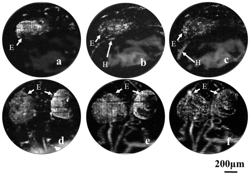
Vascular development in the heart and brain regions of the zebrafish embryo. (a-c): Longitudinal imaging of the heart in the embryonic zebrafish at 22, 25 and 28 hpf, respectively. (d-f): The sagittal view of the microvascular development in the brain at 36, 48 and 72 hpf, respectively. H: Heart E: Eye. |
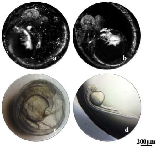
Comparison of the image quality with and without the eggshell. (a) MAP image of the embryo with the eggshell. (b) MAP image of the embryo without the eggshell. (c, d) Conventional optical microscopy of the embryos with and without the eggshell. |
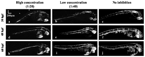
Evaluation of the pigment inhibitor. (a, b, c) MAP images of zebrafish at 30 hpf with high (a), low (b) concentrations of pigment inhibitor and without inhibition (c). (d, e, f) MAP images of the same zebrafish in (a, b, c) at 48 hpf. (e, f, j) MAP images of the same zebrafish in (a, b, c) at 60 hpf. |