
FIGURE
Fig. S2
- ID
- ZDB-FIG-171115-14
- Publication
- Fame et al., 2016 - Directional cerebrospinal fluid movement between brain ventricles in larval zebrafish
- Other Figures
- All Figure Page
- Back to All Figure Page
Fig. S2
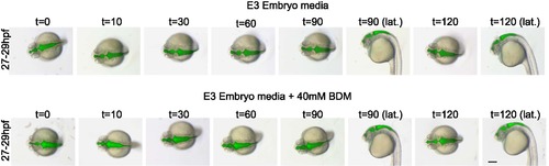
BDM treatment does not disrupt gross ventricular morphology over 2 hours. Larvae treated with 40mM BDM show no change in gross ventricular shape compared to untreated larvae. Brightfield and florescence microscopy (merged images shown) is shown after ventricular injection of 2nL of 2000kDa dextran FITC. BDM, 2,3 butanedione monoxime ; hpf, hours post-fertilization; t= time after treatment (minutes). Scale bar: 200μm. N ≥10 observed for each treatment, representative images shown. |
Expression Data
Expression Detail
Antibody Labeling
Phenotype Data
Phenotype Detail
Acknowledgments
This image is the copyrighted work of the attributed author or publisher, and
ZFIN has permission only to display this image to its users.
Additional permissions should be obtained from the applicable author or publisher of the image.
Full text @ Fluids Barriers CNS