
Fig. 2
- ID
- ZDB-FIG-171115-10
- Publication
- Fame et al., 2016 - Directional cerebrospinal fluid movement between brain ventricles in larval zebrafish
- Other Figures
- All Figure Page
- Back to All Figure Page
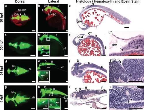
Zebrafish brain ventricle anatomy at early and late larval stages. Using 2 nL rhomencephalic ventricular injections of fluorescein-labeled dextran and imaging by Selective Plane Illumination Microscopy (SPIM, or Lightsheet Microscopy), ventricular system anatomy, volume, and connectivity was visualized in three dimensions. a Dorsal, b lateral view, 30 hpf zebrafish larvae with neuroepithelium labeled in red (mApple-caax) and the ventricular system labeled in green (fluorescein dextran injection). c H&E stain of 4 μm thick parasagittal section of 30 hpf zebrafish. aʹ Dorsal, bʹ Lateral view, 30 hpf larval zebrafish ventricular system. bʹ inset Magnification of boxed region in bʹ to show spinal canal (S) branching from the ventral aspect of the D/M ventricle (arrow). cʹ H&E stain of 4 μm thick section of 30 hpf zebrafish. cʹʹ magnification of boxed region in cʹ to show spinal canal (S) branching from D/M ventricle. d Dorsal, e Lateral view, 54 hpf larval zebrafish ventricular system. e inset Magnification of boxed region in d to show SC branching from the ventral aspect of the diencephalon (arrow). f H&E stain of 4 μm thick section of 54 hpf zebrafish. fʹ magnification of boxed region in f to show spinal canal (S) branching from diencephalon. g Dorsal, h Lateral view of the 5 dpf larval zebrafish ventricular system. h insets Magnification of boxed regions in h to show SC branching from the ventral aspect of the diencephalon (arrow) and rhombencephalon (arrow). i H&E stain of 4 μm thick section of 5 dpf zebrafish. iʹ magnification of boxed region in i to show spinal canal (S) branching from rhombencephalon. T telencephalic ventricle, D/M diencephalic/mesencephalic ventricle, D diencephalic ventricle, Te tectal ventricle, R rhombencephalic ventricle, S spinal canal, hpf hours post-fertilization, dpf days post-fertilization. Scale bar: bʹ, e, h insets, cʹʹ, fʹ, iʹ: 50 μm; all others: 100 μm |