
FIGURE
Fig. S1
- ID
- ZDB-FIG-171115-13
- Publication
- Fame et al., 2016 - Directional cerebrospinal fluid movement between brain ventricles in larval zebrafish
- Other Figures
- All Figure Page
- Back to All Figure Page
Fig. S1
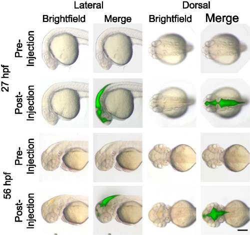
Ventricle injections do not disrupt gross ventricular morphology. Larvae were imaged under brightfield (first and third columns) and fluorescence (second and fourth columns, merged with brightfield) microscopy both before and after ventricular injection. Injections comprised 2nL of 2000kDa dextran FITC at 27 hpf (top) and 56 hpf (bottom). No change in ventricular morphology is observed in either dorsal (first two columns) or lateral (last two columns) views. hpf, hours post-fertilization. Scale bar: 200μm. N ≥10 observed at each stage, representative images shown. |
Expression Data
Expression Detail
Antibody Labeling
Phenotype Data
Phenotype Detail
Acknowledgments
This image is the copyrighted work of the attributed author or publisher, and
ZFIN has permission only to display this image to its users.
Additional permissions should be obtained from the applicable author or publisher of the image.
Full text @ Fluids Barriers CNS