
Fig. 5
- ID
- ZDB-FIG-171110-3
- Publication
- Hlushchuk et al., 2016 - Zebrafish Caudal Fin Angiogenesis Assay-Advanced Quantitative Assessment Including 3-Way Correlative Microscopy
- Other Figures
- All Figure Page
- Back to All Figure Page
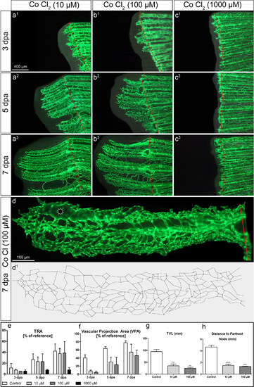
Assessment of effects of systemic cobalt chloride application at different concentrations 10, 100 and 1000μM. a-c Fluorescent images of regenerating caudal fins at 3 (a1-c1), 5 (a2-c2) and 7 dpa (a3-c3) under 3 different conditions: 10 μM (left column), 100 μM (middle) and 1000 μM of cobalt chloride (right column). Based on the high-magnification images of the single fin rays at 7 dpa (d), the skeletons of the regenerating vasculature at 7dpa were obtained (d’). The commonly used assessment values revealed no statistically significant differences (e-f). To focus on the effects of CoCl2-treatment in all applied concentrations, the controls are not displayed in this figure. The control fish used for the quantifications shown in the graphs (e-h) are the same as in Fig 4. TRA in 10- and 100 μM groups had no remarkable difference in comparison with the control values (e). At 7dpa, the vascular projection area could reveal some tendency to dose-dependent inhibition, but the difference was not statistically significant. That can be, at least, partially explained by the sporadic presence of dilated vessels (marked with an asterisk in d). The skeleton-derived parameters like total vascular length (TVL) (g) or distance to farthest node (h) are not influenced by dilated vessels and revealed statistically significant differences. *—p<0.05; **—p<0.01; ***—p<0.001; ****—p<0.0001. Remark: The mortality in the 1000μM-group was very high (only 2 out of 8 fishes survived till day 7)–that was the reason for not considering it for the advanced analysis. |