
Fig. 4
- ID
- ZDB-FIG-171110-2
- Publication
- Hlushchuk et al., 2016 - Zebrafish Caudal Fin Angiogenesis Assay-Advanced Quantitative Assessment Including 3-Way Correlative Microscopy
- Other Figures
- All Figure Page
- Back to All Figure Page
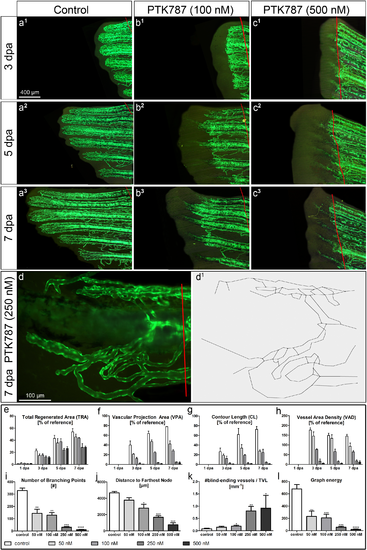
DAA: Assessment of the anti-angiogenic effects of PTK787 at different concentrations 50, 100, 250, 500 nM: a-c Fluorescent images of regenerating caudal fins at 3 (a1-c1), 5 (a2-c2) and 7 dpa (a3-c3) under 3 different conditions: vehicle control (left column), 100 nM (middle) and 500 nM PTK787 (right column). PTK787 clearly inhibits regenerative vascular outgrowth in a dose-dependent manner as confirmed by the quantitative assessment (see graphs e-l). Based on the high-magnification images of the single fin rays at 7 dpa (example in d), the skeletons of the regenerating vasculature were obtained (d1). Advanced analysis of the vasculature of the 4th ray at 7 dpa allows quantitative differentiation between multiple concentrations of PTK787. Although tissue regeneration was remarkably inhibited at 5 and 7 dpa at 250 and 500 nM (c2, c3 and e): at 50 and 100 nM of PTK787—no such prominent effects were observed (b2, b3 and e). VPA (f), CL (g) and VAD (h) showed dose-dependent inhibition with the difference between two lowest concentrations decreasing towards day 7. Four advanced (skeleton-based) parameters are presented in the lower row of graphs (i-l). Number of BPs (i) clearly distinguishes between lower and higher concentrations of PTK787, with the distance to the farthest node (j) being most informative/reliable for differentiation between all four concentrations applied. The number of blind-ending vascular segments per total vascular length (k) turned out to be capable of distinguishing the 4 concentrations applied. Interestingly, the newly introduced parameter graph energy (l) describing the connectivity of the vascular network detected highly significant differences between all the applied concentrations and the reference. *—p<0.05; **—p<0.01; ***—p<0.001; ****—p<0.0001. |