
FIGURE
Fig. 2
Fig. 2
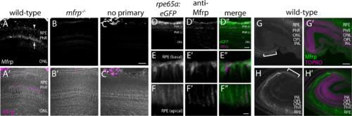
Zebrafish Mfrp is localized to the apical and basal RPE and ciliary region. (A, B, C) An anti-Mfrp antibody raised in mouse detected Mfrp in zebrafish apical and basal RPE (arrow, arrowhead, respectively). (A', B', C') Immunofluorescent signal in (A–C) is pseudocolored magenta to contrast with superimposed phase images of the cryosections. Note that rod outer segments (PhR) are found in the interval between basal and apical RPE. Cone inner and outer segments (not labeled) are in the interval between PhR and the ONL. No staining was seen when control cryosections were processed without anti-Mfrp primary antibody. Mfrp protein was seen in wild-type zebrafish RPE, but not in age-matched mutant (mfrp−/−) zebrafish with homozygous 5 bp deletions in mfrp (B, B'). Bright staining behind the eye is nonspecific staining by the secondary antibody as seen in (C). (D) Low-power image similar to (A) that includes RPE along with higher power images of basal RPE (E) and apical RPE (F) from transgenic fish expressing eGFP under control of the rpe65a promoter to delineate the RPE. Mfrp staining (D', E', F') in the RPE labels the same cells as those filled with cytoplasmic eGFP (D, E, F). Mfrp staining was seen on both apical (E') and basal (F') RPE. (D”, E”, F'') are merged images of eGFP (green) and MFRP (magenta). Note that because of the very high eGFP signal compared with that for Mfrp, (E) and (F) are single Z-slices of eGFP, whereas (E') and (F') are Z-projections through the same cells of Mfrp staining. (G, H) Mfrp staining was also seen in both the dorsal (G) and ventral (H) ciliary epithelium (brackets), contiguous with the RPE. In (G') and (H'), the Mfrp immunofluorescence is shown in green as an overlay with nuclei labeled with TO-PRO-3 (magenta). Scale bars: (A–D) 100 μm; (E–F) 20 μm; (G–H) 100 μm. PhR, photoreceptors (r, rods; c, cones); ONL, outer nuclear layer; OPL, outer plexiform layer; INL, inner nuclear layer.
|
Expression Data
| Genes: | |
|---|---|
| Fish: | |
| Anatomical Terms: | |
| Stage: | Adult |
Expression Detail
Antibody Labeling
Phenotype Data
| Fish: | |
|---|---|
| Observed In: | |
| Stage: | Adult |
Phenotype Detail
Acknowledgments
This image is the copyrighted work of the attributed author or publisher, and
ZFIN has permission only to display this image to its users.
Additional permissions should be obtained from the applicable author or publisher of the image.
Full text @ Invest. Ophthalmol. Vis. Sci.