
FIGURE
Fig. 4
Fig. 4
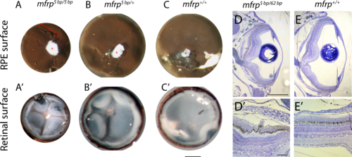
Zebrafish mfrp mutant eyes show RPE folds and compressed retinas. (A, B, C) Dissected eyecups (RPE + retina, dissected after whole-eye fixation) from mfrp mutants are smaller than sibling controls. Mutants of mfrp show folds in the RPE (red bracket), and retinas show more involutions due to compression within a smaller sclera. Optic nerves are highlighted with red asterisks. (D, E) Semithin plastic histologic sections show that mfrp mutant eyes are smaller than control siblings. RPE folds can be seen clearly at higher magnifications. Scale bars: (A–C) 1 mm; (D–E) 500 μm; (D', E') 20 μm.
|
Expression Data
Expression Detail
Antibody Labeling
Phenotype Data
| Fish: | |
|---|---|
| Observed In: | |
| Stage: | Adult |
Phenotype Detail
Acknowledgments
This image is the copyrighted work of the attributed author or publisher, and
ZFIN has permission only to display this image to its users.
Additional permissions should be obtained from the applicable author or publisher of the image.
Full text @ Invest. Ophthalmol. Vis. Sci.