
FIGURE
Fig. 5
Fig. 5
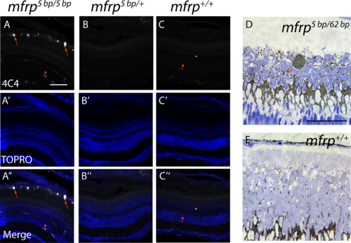
Zebrafish mfrp mutants show subretinal microglia/macrophages. (A, B, C) Cells in the subretinal space of mfrp mutant zebrafish stain positively with a microglia/macrophage marker (antibody 4C4; red arrows), whereas control heterozygous and wild-type sibling zebrafish lack such staining in the subretinal space. Resident microglia in the inner retina also stain with this marker (red arrowheads). (D, E) A pigmented subretinal cell, likely a macrophage, is visible among the photoreceptor outer segments in an mfrp mutant plastic section (red arrow). These pigmented cells are not seen in controls. Scale bars: (A–F) 100 μm; (G–H) 20 μm.
|
Expression Data
| Antibody: | |
|---|---|
| Fish: | |
| Anatomical Term: | |
| Stage: | Adult |
Expression Detail
Antibody Labeling
Phenotype Data
Phenotype Detail
Acknowledgments
This image is the copyrighted work of the attributed author or publisher, and
ZFIN has permission only to display this image to its users.
Additional permissions should be obtained from the applicable author or publisher of the image.
Full text @ Invest. Ophthalmol. Vis. Sci.