
Fig. 4
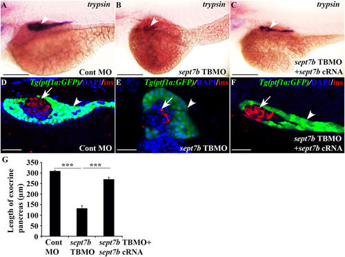
Depletion of sept7b leads to downregulation of exocrine marker trypsin and abnormal exocrine morphology. (A,B) Expression of trypsin in exocrine pancreas (arrowhead) is reduced in 3 dpf sept7b knockdown larvae (B) compared to control MO-injected larvae (A). (C) Co-injection of sept7b cRNA with sept7b TBMO restores trypsin expression (arrowhead). (D,E) sept7b TBMO-injected Tg(ptf1a:GFP) zebrafish larvae (E) show abnormal exocrine morphology and reduced size of the pancreatic tail (arrowhead) compared to the control MO-injected larvae (D) at 3 dpf. β-cells are labeled with insulin (red; arrow). (F) Co-injection of sept7b cRNA with sept7b TBMO partially restores the morphology of exocrine pancreas. In (D–F), nuclei are labeled with DAPI (blue). (G) The length of the exocrine pancreas, measured from 3 dpf zebrafish embryos using trypsin as a marker, is significantly decreased by sept7b knockdown compared to the control. Co-injection of sept7b cRNA with sept7b TBMO restores the length of the exocrine pancreas. Scale bar: (A–C) (75 μm); (D–F) (25 μm). Error bars represent mean ± SEM. ***p ≤ 0.0005. |
| Genes: | |
|---|---|
| Antibody: | |
| Fish: | |
| Knockdown Reagent: | |
| Anatomical Terms: | |
| Stage: | Protruding-mouth |
| Fish: | |
|---|---|
| Knockdown Reagent: | |
| Observed In: | |
| Stage: | Protruding-mouth |