
Fig. 3 S1
- ID
- ZDB-FIG-170609-66
- Publication
- Gong et al., 2017 - The Sec14-like phosphatidylinositol transfer proteins Sec14l3/SEC14L2 act as GTPase proteins to mediate Wnt/Ca2+ signaling.
- Other Figures
- All Figure Page
- Back to All Figure Page
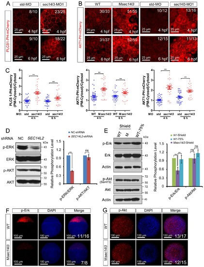
sec14l3 depletion results in PIP2 and PIP3 accumulation in the PM. (A) Accumulation of PIP2 bound by PLCδ1-PH-mCherry in the PM of sec14l3 morphants at 4 and 6 hpf. The shown pictures are immunofluorescent images of embryos viewed from the animal-pole. Scale bars, 25 μm. (B) Accumulation of PIP3 bound by AKT1-PH-mCherry in the PM of Msec14l3 mutants (the left two columns) and sec14l3 morphants (the right two columns) at 4 and 6 hpf. Scale bars, 25 μm. (C) Quantification of PM PIP2/PIP3 accumulation in embryonic cells of (A–B). The PM PIP2/PIP3 accumulation was respectively reflected by the PLCδ1/AKT1-PH-mCherry intensity ratio of (PM-Cytosol)/Cytosol. Embryos from three independent experiments were calculated (see also Figure 3—source data 1, **p<0.01). (D) Western blot analysis of phosphorylated ERK and AKT in SEC14L2 knockdown stable HEK293T cells. A notable reduction of p-ERK(Thr202/Tyr204) occurred, but no change of p-AKT(Ser473) was observed in SEC14L2 depleted cells. Statistical analysis is shown on the right. Data are presented as mean ± SEM based on three independent experiments (see also Figure 3—source data 1, **p<0.01; ns, non-significant, p>0.05). (E) Phosphorylation levels of the Erk and Akt in Msec14l3 mutant embryos. p-Erk, total Erk, p-Akt, total Akt and Actin were examined at the shield stage by western blot using corresponding antibodies. Statistical analysis is shown on the right. Data are presented as mean ± SEM based on three independent experiments (see also Figure 3—source data 1, **p<0.01; ns, non-significant, p>0.05). (F–G) Immunostaining images of p-Erk (C) and p-Akt (D) in Msec14l3 mutant embryos at the sphere stage. Nuclei were stained with DAPI. Lateral views (C) and animal-pole views (D) are shown respectively. Scale bars, 100 μm. |