
Fig. 1 S2
- ID
- ZDB-FIG-170609-63
- Publication
- Gong et al., 2017 - The Sec14-like phosphatidylinositol transfer proteins Sec14l3/SEC14L2 act as GTPase proteins to mediate Wnt/Ca2+ signaling.
- Other Figures
- All Figure Page
- Back to All Figure Page
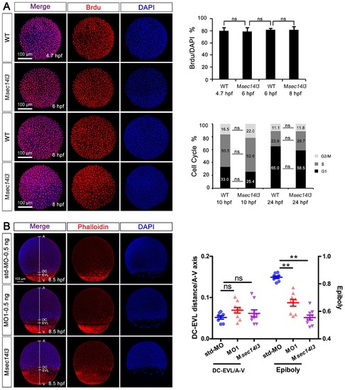
Sec14l3 depletion has no effect on cell proliferation or cell cycle progression, as well as cell adhesion between envelop layer (EVL) and deep cells (DC). (A) Cell proliferation analysis based on Brdu incorporation and cell cycle progression analyzed by flow cytometry were performed. The ratios of Brdu/DAPI indicating percentages of proliferating cells are shown at the right top; Right bottom panel shows the result of cell cycle analysis, and the percentages of individual phases are labeled. Light grey indicates G2/M phase, grey indicates S phase and black indicates G1 phase. No evident difference is observed between control and mutant embryos. Data are presented as mean ± SEM. Significance was analyzed using Student’s t-test (ns, non-significant). scale bars, 100 μm. (B) Distance measurement between envelop layer (EVL) and deep cells (DC) based on phalloidin (red) and DAPI (blue) staining, see also in Figure 1—source data 1. The left panel shows immunostaining results in sec14l3 morphants and Msec14l3 mutants at 8.5 hpf. Embryos were laterally viewed with animal pole (A) to the top, vegetal pole (V) to the bottom and the marginal borders of DC and EVL are marked. Statistical results of 10 embryos from each group are shown at the right panel. Ratios of DC-EVL distance to A-V axis and epiboly are calculated. |