
Fig. S1
- ID
- ZDB-FIG-170609-43
- Publication
- Theodore et al., 2017 - Distinct Roles for Matrix Metalloproteinases 2 and 9 in Embryonic Hematopoietic Stem Cell Emergence, Migration, and Niche Colonization
- Other Figures
- All Figure Page
- Back to All Figure Page
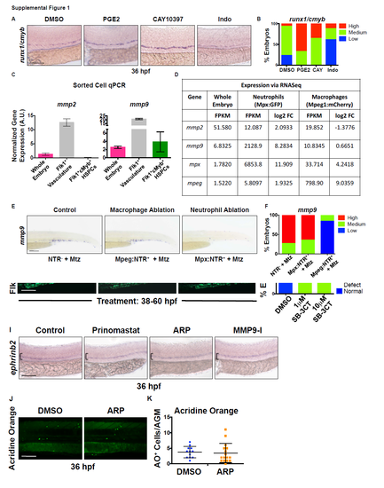
Mmp2 and Mmp9 are expressed in discrete populations within hemogenic regions. Related to Figure 1.
A) Exposure to dimethyl-Prostaglandin-E2 (PGE2) (10μM) and CAY10397 (CAY) (10μM), a small molecule that stabilizes endogenous PGE2, increased runx1 and cmyb expression in the ventral dorsal aorta (VDA) by WISH, while the cyclooxygenase antagonist Indomethacin (Indo, 10μM) decreased expression of HSPC markers at 36 hours post fertilization (hpf). B) Qualitative phenotypic distribution of embryos from S1A scored with high/medium/low runx1/cmyb expression in the AGM (n≥20 embryos/condition). C) qPCR analysis using FACS-sorted cell populations from Tg(kdrl:dsred;cmyb:gfp) embryos at 36 hpf showed enrichment of mmp2 in the Flk1+cMyb- vasculature, while mmp9 expression was present in all populations examined (1000 embryos x 2 replicate sorts). Error bars: mean ± SD. D) RNA-seq analysis (~1000 pooled Tg(mpeg1:mcherry;mpx:egfp) embryos, >50,000 cells/fraction) indicated that mmp9 expression is concentrated in FACS-sorted Mpx+ neutrophils at 72 hpf. E) Metronidozole (Mtz)-mediated (24-48 hpf) targeted ablation of primitive myeloid cell types in Tg(mpeg1:gal4;uas:nfsb-mcherry) (macrophage) or Tg(mpx:gal4;uas:nfsb-mcherry) (neutrophil) embryos confirmed that neutrophil loss strongly diminished mmp9 expression in the trunk and tail, while macrophage loss had minimal impact. F) Qualitative phenotypic distribution of embryos from S1E scored with high/medium/low mmp9 expression in the trunk and tail region (n≥25 embryos/condition). Scale bars: 100μm. |