
Fig. 6
- ID
- ZDB-FIG-170609-12
- Publication
- Kelu et al., 2017 - Ca2+ release via two-pore channel type 2 (TPC2) is required for slow muscle cell myofibrillogenesis and myotomal patterning in intact zebrafish embryos.
- Other Figures
- All Figure Page
- Back to All Figure Page
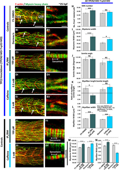
Effect of IP3/BM and caffeine on the organization of the trunk musculature and the formation of the sarcomeres after TPC2-knockdown. Embryos were (A-E) injected with TPCN2-MO-T and p53-MO at the 1-cell stage or (F,G) allowed to develop without MO injection. At 17 hpf, the terminal portion of the tail was excised after which embryos were treated with either: (A) 1% DMSO in Danieau's solution (control of the IP3/BM treatment); (B) Danieau's solution alone (control of the caffeine treatment), (C,F) IP3/BM at 100 µM; or (D,E,G) caffeine at (D) 0.5 mM or (E,G) 2 mM. Embryos were then fixed at ~25 hpf, and dual-labeled with phalloidin and the F59 antibody, to visualize F-actin (in red) and myosin heavy chain (in green) in the trunk musculature, respectively. The panels represent a series of optical sections projected as single images at (Ai-Gi) low and (Aii-Gii) higher magnification when the red and green channels are merged; overlapping regions are shown in yellow. The higher magnification images of the SMC myofibers reveal the presence or absence of the sarcomeric banding pattern of the F-actin and myosin heavy chain. The white arrows in panels Ai, Bi, Di and Ei, show examples of elongated SMC myofibers. Ant. and Pos. in panel Ai are anterior and posterior, respectively. Scale bars, 50 µm in panels Ai-Gi; and 2 µm, in panels Aii-Gii. (H-L) Quantification of the effect of IP3/BM and caffeine after TPC2-knockdown on the organization of the trunk musculature and the formation of the sarcomeres. These bar charts show the mean ±SEM: (H) number of SMCs; (I) myotome width, and (J) somite angle, (all n=12 from 4 embryos); as well as the (K) myofiber length: somite length ratio (these lengths were measured in n=72 myofibers from 4 embryos); and (L) myofiber width (n=40, from 4 embryos). The dashed black line in panel (K) indicates a myofiber: somite length ratio of 1. (M,N) Quantification of the effect of IP3/BM and caffeine in control embryos (without TPC2 knockdown) on (M) the myotome width (n≥9, from ≥3 embryos), and (N) myofiber width (n≥30, from ≥3 embryos). The asterisks indicate statistically differences at p<0.05 (*) and p<0.0005 (***). |
| Antibody: | |
|---|---|
| Fish: | |
| Conditions: | |
| Knockdown Reagents: | |
| Anatomical Term: | |
| Stage: | Prim-5 |
| Fish: | |
|---|---|
| Conditions: | |
| Knockdown Reagents: | |
| Observed In: | |
| Stage: | Prim-5 |
Reprinted from Developmental Biology, 425(2), Kelu, J.J., Webb, S.E., Parrington, J., Galione, A., Miller, A.L., Ca2+ release via two-pore channel type 2 (TPC2) is required for slow muscle cell myofibrillogenesis and myotomal patterning in intact zebrafish embryos., 109-129, Copyright (2017) with permission from Elsevier. Full text @ Dev. Biol.