
Fig. 5
- ID
- ZDB-FIG-170607-16
- Publication
- Kelu et al., 2017 - Ca2+ release via two-pore channel type 2 (TPC2) is required for slow muscle cell myofibrillogenesis and myotomal patterning in intact zebrafish embryos.
- Other Figures
- All Figure Page
- Back to All Figure Page
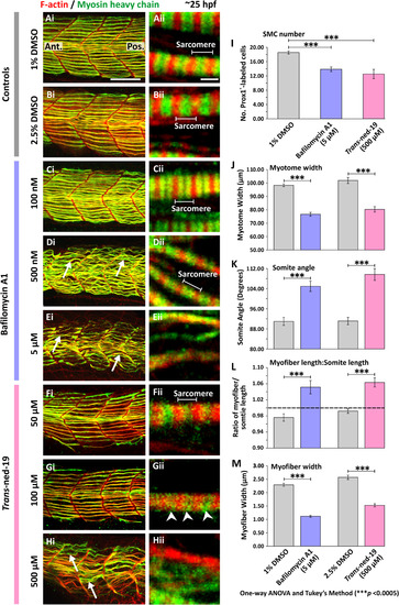
Effect of bafilomycin A1 and trans-ned-19 on the organization of the trunk musculature and the formation of the sarcomeres. At 17 hpf, embryos had the terminal portion of the tail excised and then were treated with either: (A) 1% DMSO or (B) 2.5% DMSO in Danieau's solution (controls of the bafilomycin A1 and trans-ned-19 treatment, respectively), or (C-E) bafilomycin A1 at: (C) 100 nM, (D) 500 nM, or (E) 5 µM; or (F-H) trans-ned-19 at (F) 50 µM, (G) 100 µM or (H) 500 µM. All the embryos were fixed at ~25 hpf and dual-labeled with phalloidin and the F59 antibody, to visualize F-actin (in red) and myosin heavy chain (in green) in the trunk musculature, respectively. The panels show a series of optical sections projected as single images at (Ai-Hi) low and (Aii-Hii) higher magnification when the red and green channels are merged; overlapping regions are shown in yellow. The higher magnification images of the SMC myofibers reveal the presence or absence of the sarcomeric banding pattern of the F-actin and myosin heavy chain. The white arrows in panels Di, Ei and Hi show examples of elongated SMC myofibers; and the white arrowheads in panel Gii indicate the appearance of some banding but clear sarcomeres are not obvious. Ant. and Pos. in panel Ai are anterior and posterior, respectively. Scale bars, 50 µm in panels Ai-Hi; and 2 µm, in panels Aii-Hii. (I-M) Quantification of the effect of bafilomycin A1 and trans-ned-19 on the organization of the trunk musculature and the formation of the sarcomeres. These bar charts show the mean ±SEM: (I) number of SMCs; (J) myotome width, and (K) somite angle, (all n=12 from 4 embryos); as well as the (L) myofiber length: somite length ratio (these lengths were measured in n=72 myofibers from 4 embryos); and (M) myofiber width (n=40, from 4 embryos). The dashed black line in panel (L) indicates a myofiber: somite length ratio of 1. The asterisks indicate statistically differences at p<0.0005 (***) |
| Antibody: | |
|---|---|
| Fish: | |
| Conditions: | |
| Anatomical Term: | |
| Stage: | Prim-5 |
| Fish: | |
|---|---|
| Conditions: | |
| Observed In: | |
| Stage: | Prim-5 |
Reprinted from Developmental Biology, 425(2), Kelu, J.J., Webb, S.E., Parrington, J., Galione, A., Miller, A.L., Ca2+ release via two-pore channel type 2 (TPC2) is required for slow muscle cell myofibrillogenesis and myotomal patterning in intact zebrafish embryos., 109-129, Copyright (2017) with permission from Elsevier. Full text @ Dev. Biol.