
Fig. 1
- ID
- ZDB-FIG-170530-9
- Publication
- Wan et al., 2017 - Opposing Actions of Fgf8a on Notch Signaling Distinguish Two Muller Glial Cell Populations that Contribute to Retina Growth and Regeneration
- Other Figures
- All Figure Page
- Back to All Figure Page
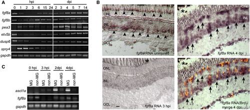
fgf8a Gene Regulation during Retina Regeneration (A) RT-PCR analysis of fgf8a, fgf8b, and Fgf-responsive genes in uninjured and needle-poke-injured retinas. (B) In situ hybridization assays and BrdU immunofluorescence for fgf8a expression and MG proliferation, respectively, before and after injury to central retina in 6-month-old fish. Arrowheads point to fgf8a RNA in uninjured retina; arrows point to fgf8a RNA enriched in proliferating MG-derived progenitors at 4 dpi. Asterisk indicates injury site. Scale bar, 50 μm. (C) RT-PCR analysis of ascl1, fgf8a, and gapdh RNAs in GFP+ MGs and GFP− non-MGs (retinal neurons) in uninjured and injured retinas that were FACS purified from gfap:GFP transgenic fish retinas. See also Figure S1. |