
FIGURE
Fig. 8
- ID
- ZDB-FIG-170522-14
- Publication
- Xavier et al., 2017 - Comparative analysis of monoaminergic cerebrospinal fluid-contacting cells in Osteichthyes (bony vertebrates)
- Other Figures
- All Figure Page
- Back to All Figure Page
Fig. 8
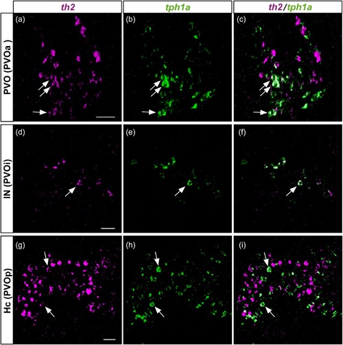
Colocalization of th2 and tph1a in the three CSF-c cell populations of zebrafish. Frontal sections of the adult zebrafish brain showing the three CSF-c cell populations, PVO (a–c), IN (d–f), and Hc (g–i), which are also known as PVOa, PVOi, and PVOp, respectively. Single confocal planes show that th2 (magenta) and tph1a (green) are found in the same cell population, and some cells coexpress both of them (arrows). Scale bar = 20 µm |
Expression Data
Expression Detail
Antibody Labeling
Phenotype Data
Phenotype Detail
Acknowledgments
This image is the copyrighted work of the attributed author or publisher, and
ZFIN has permission only to display this image to its users.
Additional permissions should be obtained from the applicable author or publisher of the image.
Full text @ J. Comp. Neurol.