
Fig. 1
- ID
- ZDB-FIG-170522-12
- Publication
- Xavier et al., 2017 - Comparative analysis of monoaminergic cerebrospinal fluid-contacting cells in Osteichthyes (bony vertebrates)
- Other Figures
- All Figure Page
- Back to All Figure Page
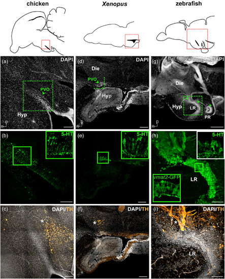
Monoaminergic CSF-c cells of chicken, Xenopus, and zebrafish in relation to the organization of the hypothalamic ventricle. Schematic drawings of sagittal sections of chicken, Xenopus, and zebrafish brains are shown in the top panels, and for each species, micrographs of the red squared areas are shown below (a–c for chicken, d–f for Xenopus, and g–i for zebrafish). DAPI staining (gray) delineates the recesses of the hypothalamic ventricle. Confocal images (Z-projection = 10 µm) with higher magnification obtained from the area depicted in (a) (dashed green square) show 5-HT+ CSF-c cells (green) aligned along the dorsal side of the hypothalamic recess (b; inset at higher magnification). TH immunoreactive cells (orange) are not observed within the PVO (arrowhead), but are abundant in the area dorsocaudal to it (c; asterisk). In the Xenopus sagittal section close to the midline, PVO (d; arrowhead) is observed at the anterior edge of the large ventricle (v). The PVO is visualized with 5-HT+ CSF-c cells (e; green; inset at higher magnification). TH immunoreactive cells (orange) are observed dorsal to the PVO (f; asterisk). In zebrafish, three CSF-c cell populations (locations indicated by arrowheads in g) are located around two hypothalamic recesses. The two anterior CSF-c cell populations are located in front of and around the lateral recess (LR), while the posterior population surrounds the posterior recess (PR). Higher magnification of the squared area in (g) is shown in (h) and (i) (Z-projection = 10 µm). CSF-c cells revealed by the expression of GFP in the enhancer trap transgenic line ETvmat2:GFP (vmat2-GFP; green inset) are lined along the ventricular zone (h). The white inset in (h) shows the 5-HT labeling in the same area (the image is taken from a different sample). TH immunoreactive cells (orange) are found dorsal to the LR (i; asterisk). D = dorsal; Die = diencephalon; Hyp = hypothalamus; LR = lateral recess; PR = posterior recess; PVO = paraventricular organ; R = rostral; v = ventricle. Scale bar = 200 µm in (a–g); 50 µm in (h, i) |